Arizona Ultrasound
@SonoStache
This is the official twitter feed for all things Arizona and Emergency Ultrasound! Program Director: Dr. Srikar Adhikari
ID:2559959808
http://emergencymed.arizona.edu/fellowships/emergency-ultrasound 10-06-2014 21:47:59
488 Tweets
3,0K Followers
172 Following
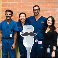
 on Twitter photo 2023-10-09 19:44:38 Fellowship friends ❤️ circa early 2000s #ACEP23")
(@SAEMAEUS) 's Twitter Profile Photo")
 (@SAEMAEUS) on Twitter photo 2023-01-30 16:00:33 Nominations are now open for the #SAEM AEUS Academy awards! Nominate yourself or someone you admire for this honor. Awards will be presented at the #SAEM23 Annual Meeting. Nominate now: bit.ly/3vweblE")
Today's #SonoGames22 Round 0 Infographic comes from Yes_We_SCAN and reviews an important paper by researchers from The University of Arizona EM Residency, MGH Emergency Med, & University of Sydney! Hamid Shokoohi Arizona Ultrasound Sid Patanwala Richard Amini
 (@SAEMAEUS) on Twitter photo 2023-01-02 17:00:01 Today's #SonoGames22 Round 0 Infographic comes from @scan_yes and reviews an important paper by researchers from @UArizonaEM, @MassGeneralEM, & @Sydney_Uni! @HamidShokoohiMD @SonoStache @sidpatan @UofASono")
 's Twitter Profile Photo")
 on Twitter photo 2022-09-28 16:01:55 Development and Validation of a Point-of-Care-Ultrasound Image Quality Assessment Tool: The POCUS IQ Scale pubmed.ncbi.nlm.nih.gov/36165271/")
 on Twitter photo 2022-09-27 03:40:38 The commemorative cheese plate has arrived! @SCUFellowships #SCUF22")
 on Twitter photo 2022-09-08 05:07:47 When past fellows unite. Class of 2021 and 2022👬🏼")
 on Twitter photo 2022-08-11 05:25:46 Our fellows love our residents… a lot")
 on Twitter photo 2022-06-28 01:18:31 Graduation night! Congrats to our fellows🌟(Bonus: a maskless Srikar) @rmohty @DAH__313")
 on Twitter photo 2022-06-13 18:12:46 One of our fellows passed his RDCS! 🙌🏽 @rmohty (FYI: He took it for fun)")
 on Twitter photo 2022-05-11 23:19:26 Another epic debate at #SAEM22 @TeddyDanielz @Takeokun & @AlTiradoUS")
 on Twitter photo 2022-05-11 21:41:38 The debate is on at #SAEM22 ! #POCUS for nec fasc?")
 on Twitter photo 2022-05-11 20:21:31 Packed house for @DAH__313 (aka DJ dispo!!!) and his Delphi study! Hard work pays off. Looking forward to the pub #SAEM22")