Rubén Tesoro Moreno(@TesoroRuben) 's Twitter Profile Photo

Sunday render!

How cells control these stunning shapes is fascinating 🤯. Can you guess which direction is moving?

*MDAMB231 cells with a membrane marker, imaged with confocal*

account_circle
JExpMed(@JExpMed) 's Twitter Profile Photo

Our May Issue is out! ➡️ hubs.la/Q02vLmwy0
The cover shows a confocal image of mouse proximal colon with epithelial cells shown in blue. From Mohammad Arifuzzaman and colleagues (hubs.la/Q02vLhhv0).

Our May Issue is out! ➡️ hubs.la/Q02vLmwy0
The cover shows a confocal image of mouse proximal colon with epithelial cells shown in blue. From @arif_uzz_aman and colleagues (hubs.la/Q02vLhhv0).
account_circle
onça-mãe(@sniperdosertao) 's Twitter Profile Photo

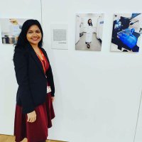
Posso não ser tão bonita a ponto de ser modelo, mas meus filhos são 😜🥰 um deles virou garoto propaganda do laboratório de microscopia confocal da USP de Ribeirão 💃🏼❤️

Posso não ser tão bonita a ponto de ser modelo, mas meus filhos são 😜🥰 um deles virou garoto propaganda do laboratório de microscopia confocal da USP de Ribeirão 💃🏼❤️
account_circle
A. I.(@MacroWorld21) 's Twitter Profile Photo

For nerds who likes to know more about Rahman confocal laser microscope using pinhole! And spectroscopy. 😛
edinst.com/blog/what-is-c…

For nerds who likes to know more about Rahman confocal laser microscope using pinhole! And spectroscopy. 😛
edinst.com/blog/what-is-c…
account_circle
HORIBA Scientific(@HScientific) 's Twitter Profile Photo

🚀 We are thrilled to unveil our latest innovation: the new laser scanning confocal FLIM platform. 🌟 This cutting-edge addition perfectly complements our existing range of HORIBA FLIM solutions, promising exciting possibilities for our customers.

account_circle
Irem Denizli(@IremDenizliMSc) 's Twitter Profile Photo

Back from the break and listening Nicola Smylie’s talk about quantitative analysis of endogenous clock proteins in mammalian SCN by confocal microscopy! BioClocksUK Laboratory of Seasonal Biology

Back from the break and listening Nicola Smylie’s talk about quantitative analysis of endogenous clock proteins in mammalian SCN by confocal microscopy! @BioClocksUK @Stevenson_Lab
account_circle
BarrowNeurological(@BarrowNeuro) 's Twitter Profile Photo

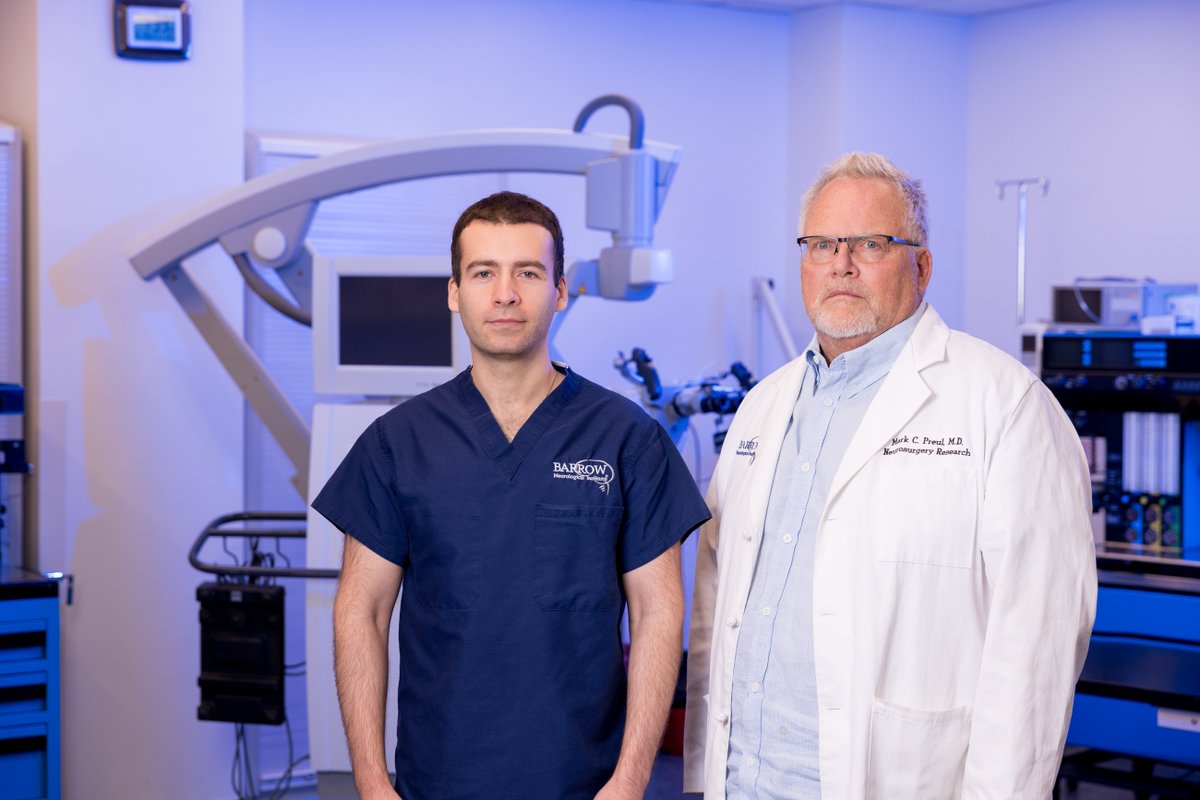
Congrats to research fellow Dr. Irakliy Abramov on receiving the Zeiss Award from AANS! He'll present his abstract, “Intraop. confocal laser endomicroscopy during resection of low-grade & nonenhancing in eloquent brain region,” at .

Congrats to research fellow Dr. Irakliy Abramov on receiving the Zeiss #BrainTumorResearch Award from @AANSNeuro! He'll present his abstract, “Intraop. confocal laser endomicroscopy during resection of low-grade & nonenhancing #braintumors in eloquent brain region,” at #AANS2024.
account_circle
ISTCT(@ISTCT_Caen) 's Twitter Profile Photo


📰 New publication
Combining sophisticated fast FLIM, confocal microscopy, ans STED nanoscopy for live-cell imaging of tunneling nanotubes by Bénard et al. in Life Science Alliance
▶ doi: 10.26508/lsa.202302398

#tunnelingnanotubes #fluorescence #cells #microscopy
📰 New publication
Combining sophisticated fast FLIM, confocal microscopy, ans STED nanoscopy for live-cell imaging of tunneling nanotubes by Bénard et al. in Life Science Alliance
▶ doi: 10.26508/lsa.202302398
account_circle
Mary Brown(@GopherConfocal) 's Twitter Profile Photo

Recognising the importance and impact of Imaging Scientists: Global guidelines for establishing career paths within core facilities - Wright - Journal of Microscopy - Wiley Online Library onlinelibrary.wiley.com/doi/10.1111/jm…

account_circle
Mahindra University Centre for Life Sciences(@CLS4MU) 's Twitter Profile Photo

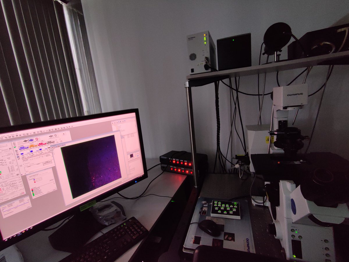
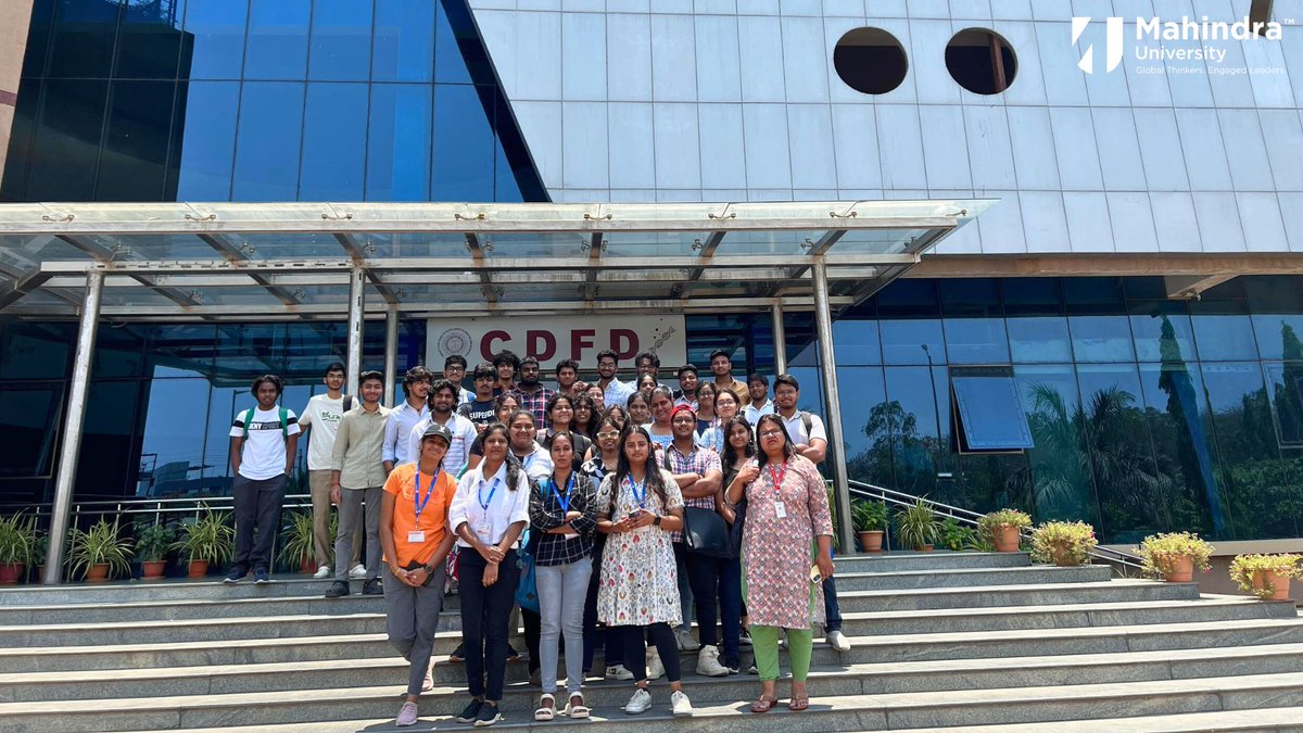
𝐃𝐫. 𝐀𝐤𝐚𝐧𝐤𝐬𝐡𝐚 𝐒𝐢𝐧𝐠𝐡 organized a scientific visit CDFD on April 24th, 2024. During this visit, students explored the SEF, where scientists demonstrated and explained the use of advanced instruments such as circular dichroism, Confocal Microscopy, FACS, and FPLC.

𝐃𝐫. 𝐀𝐤𝐚𝐧𝐤𝐬𝐡𝐚 𝐒𝐢𝐧𝐠𝐡 organized a scientific visit CDFD on April 24th, 2024. During this visit, students explored the SEF, where scientists demonstrated and explained the use of advanced instruments such as circular dichroism, Confocal Microscopy, FACS, and FPLC.
account_circle
Fabrizio Mattei(@FabriMattei) 's Twitter Profile Photo

Glad to present this methodological preprint showing how Confocal Microscopy station can be coupled to organ-on-chip platforms to precisely determine 3D spatial distribution between immune cells and tumor cells upon epigenetic drug treatment.
biorxiv.org/content/10.110…

account_circle
Biological Optical Microscopy Platform(@BOMPUoM) 's Twitter Profile Photo

We have an opening for an applications specliast to join our team. You will use your microscopy expertise to work collaboratively with researchers using advanced applications on platform equipment including Lightsheet and confocal systems.

account_circle
Nico Schramma(@nicoschramma) 's Twitter Profile Photo

So actually, the chloroplast is auto fluorescent, which allowed us to do some confocal microscopy under blue light stimulation.
We discover that it's not a gooey gel, but a reticulated cytoplasmic network. That can buckle rapidly into the free space between the strands.

8/12

account_circle