Terkild Brink Buus(@TerkildB) 's Twitter Profile Photo

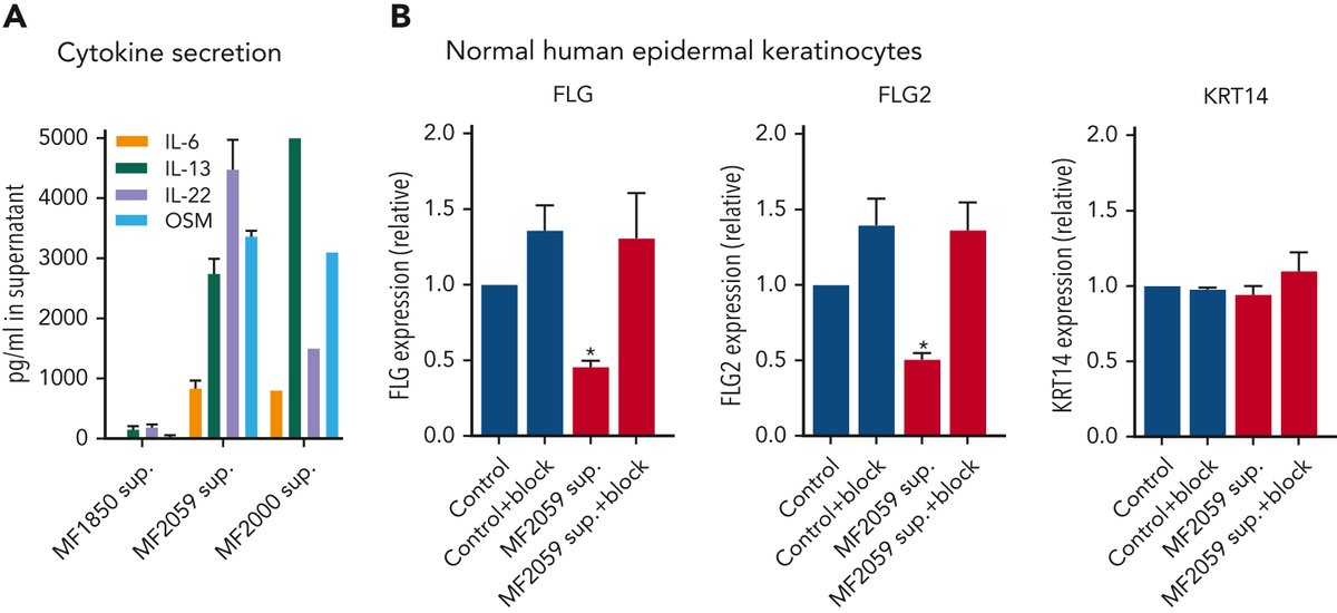
Several cytokines can repress FLG through JAK1/STAT3 signaling in keratinocytes. We found high levels of IL-6, IL-13, IL-22 and OSM secreted by malignant T cells. Blocking these completely counteracted the effect of malignant T cells on FLG expression in keratinocytes.

Several cytokines can repress FLG through JAK1/STAT3 signaling in keratinocytes. We found high levels of IL-6, IL-13, IL-22 and OSM secreted by malignant T cells. Blocking these completely counteracted the effect of malignant T cells on FLG expression in keratinocytes.
account_circle
🥼Agingdoc1⭐MD, PhD 🔔(@agingdoc1) 's Twitter Profile Photo

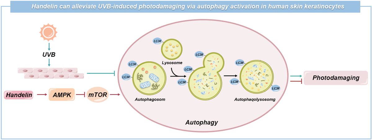
Handelin protects human skin keratinocytes against ultraviolet B-induced photodamage via autophagy activation by regulating the AMPK-mTOR signaling pathway

sciencedirect.com/science/articl…

Handelin protects human skin keratinocytes against ultraviolet B-induced photodamage via autophagy activation by regulating the AMPK-mTOR signaling pathway

sciencedirect.com/science/articl…
account_circle
Eve Examines👩🏻‍🦼🇺🇦 🔸🐾🦨🦥🌷🪻🦦🦤🪺(@EveExamines) 's Twitter Profile Photo

Cindy Lu Jette is Mataray 😊 Thank you. Yeah that's what I thought, we know it's an auto immune process. It's a viral induced autoimmune disease.

Was reading about psoriasis yesterday. The keratinocytes make chemicals that keep the T cells attacking them. Like a hellish feedback loop.

account_circle
Terkild Brink Buus(@TerkildB) 's Twitter Profile Photo

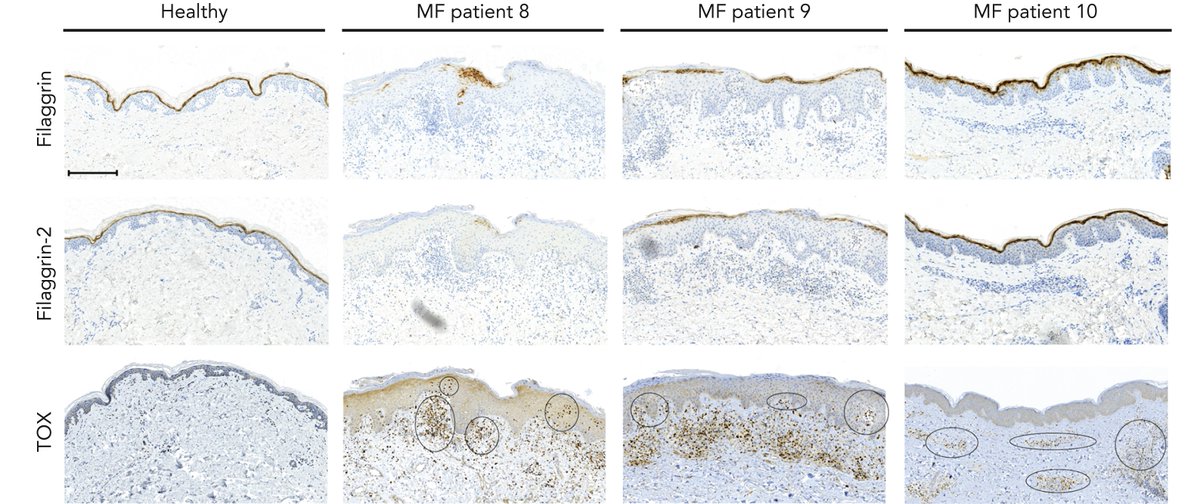
Malignant (cancer) T cells in CTCL express high levels of the TOX protein. Association of defective skin barrier and TOX+ cells close to epidermis suggest that the defect may be caused by the proximity of malignant T cells and epidermal keratinocytes.

Malignant (cancer) T cells in CTCL express high levels of the TOX protein. Association of defective skin barrier and TOX+ cells close to epidermis suggest that the defect may be caused by the proximity of malignant T cells and epidermal keratinocytes.
account_circle
Niacin + 3.5x N-AcetylGlutamine(@GPR109A) 's Twitter Profile Photo

… Supplementation of nicotinic acid and its derivatives up-regulates cellular NAD+ level rather than nicotinamide derivatives in cultured normal human epidermal keratinocytes

researchsquare.com/article/rs-248…

account_circle
Kaleb(@901Burner) 's Twitter Profile Photo

Zachary Smith Ok, Keratinocytes are the predominant cell type of epidermis and originate in the basal layer, produce keratin, and are responsible for the formation of the epidermal water barrier by making and secreting lipids.
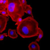
account_circle
SarcomaPapers(@Sarcoma_papers) 's Twitter Profile Photo

Effects of 5G-modulated 3.5 GHz radiofrequency field exposures on HSF1, RAS, ERK, and PML activation in live fibroblasts and keratinocytes cells dlvr.it/SpWngg

account_circle
Eve Examines👩🏻‍🦼🇺🇦 🔸🐾🦨🦥🌷🪻🦦🦤🪺(@EveExamines) 's Twitter Profile Photo

Dr. Lisa Iannattone T. Ryan Gregory Yes, if it were vindictive bureaucrats it would be unsurprising. But good gravy! They sure aren't like the authors of the paper I read the abstract of about the interaction between T cells and keratinocytes in psoriasis. Fucking amazing! So complicated.

account_circle
Bliq Photonics(@BliqPhotonics) 's Twitter Profile Photo

🔬 Two-photon excitation and CARS microscopy for structural and molecular assessment of lesions in multiple sclerosis 🔬

💡 What are your in vivo imaging challenges? bliqphotonics.com/solutions/mult…

🔬 Two-photon excitation and CARS microscopy for structural and molecular assessment of lesions in multiple sclerosis 🔬

💡 What are your in vivo imaging challenges? bliqphotonics.com/solutions/mult…

#microscopy #twophoton #multiphoton #invivo
account_circle
Terkild Brink Buus(@TerkildB) 's Twitter Profile Photo

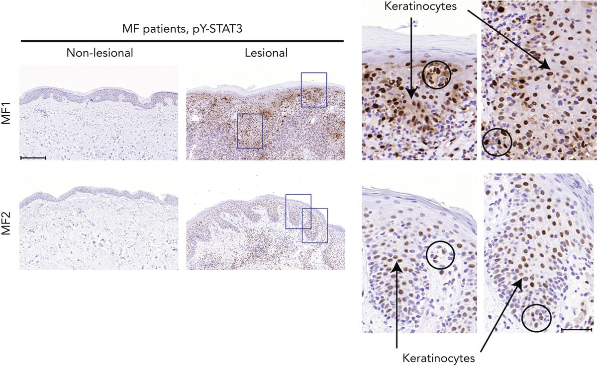
JAK/STAT3 signaling plays a key role in repression of FLG in inflammatory skin disorders but little is known about STAT3 activity CTCL skin. We found strong STAT3 activation in T cells and keratinocytes in lesional, but not in non-lesional, skin of CTCL patients.

JAK/STAT3 signaling plays a key role in repression of FLG in inflammatory skin disorders but little is known about STAT3 activity CTCL skin. We found strong STAT3 activation in T cells and keratinocytes in lesional, but not in non-lesional, skin of CTCL patients.
account_circle
Gillies McIndoe Research Institute(@Gillies_McIndoe) 's Twitter Profile Photo

Alex Chan led our this month. He & Dr Maddie White chose, “Rapid culture of human keratinocytes in an autologous, feeder-free system with a novel growth medium,” written by Feisst et al. University of Auckland | Waipapa Taumata Rau and published by Cytotherapy Journal (@ISCTglobal).

Alex Chan led our #JournalClub this month. He & Dr Maddie White chose, “Rapid culture of human keratinocytes in an autologous, feeder-free system with a novel growth medium,” written by Feisst et al. @AucklandUni and published by @cytotherapy Journal (@ISCTglobal).

#novelmethod
account_circle
Chengdu Youngshe Peptide(@AileenYYDS) 's Twitter Profile Photo

Tetrapeptide-61 is a peptide which delivers improve skin vitality and yields a younger skin appearance. This works especially on aged fibroblasts and keratinocytes to allow them to regain their youthful energy and promote anti-aging appearances.

Chengdu Youngshe Chemical Co.,Ltd

Tetrapeptide-61 is a peptide which delivers improve skin vitality and yields a younger skin appearance. This works especially on aged fibroblasts and keratinocytes to allow them to regain their youthful energy and promote anti-aging appearances.

Chengdu Youngshe Chemical Co.,Ltd
account_circle
Terkild Brink Buus(@TerkildB) 's Twitter Profile Photo

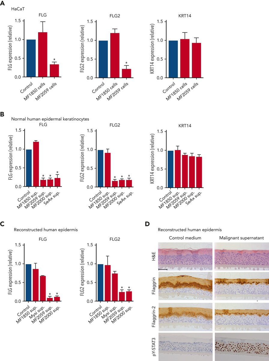
Indeed, culture of keratinocytes in the presence of malignant T cells (unlike non-malignant T cells) is sufficient to reduce the expression of barrier proteins (FLG & FLG-2). This reduction is caused by soluble mediators and associated with STAT3 activation in keratinocytes.

Indeed, culture of keratinocytes in the presence of malignant T cells (unlike non-malignant T cells) is sufficient to reduce the expression of barrier proteins (FLG & FLG-2). This reduction is caused by soluble mediators and associated with STAT3 activation in keratinocytes.
account_circle
Research Square Preprints(@RS_Preprints) 's Twitter Profile Photo

Immature mandarin orange extract increases the amount of Hyaluronic acid in human skin fibroblast and keratinocytes researchsquare.com/article/rs-281…

account_circle
Terkild Brink Buus(@TerkildB) 's Twitter Profile Photo

TLDR: Cytokine-producing malignant T cells markedly reduce barrier protein expression in keratinocytes through JAK/STAT3 signaling. Barrier protein is restored by JAK-inhibitors indicating them as a novel treatment option in CTCL patients

Highlights below
doi.org/10.1182/blood.…

account_circle
Terkild Brink Buus(@TerkildB) 's Twitter Profile Photo

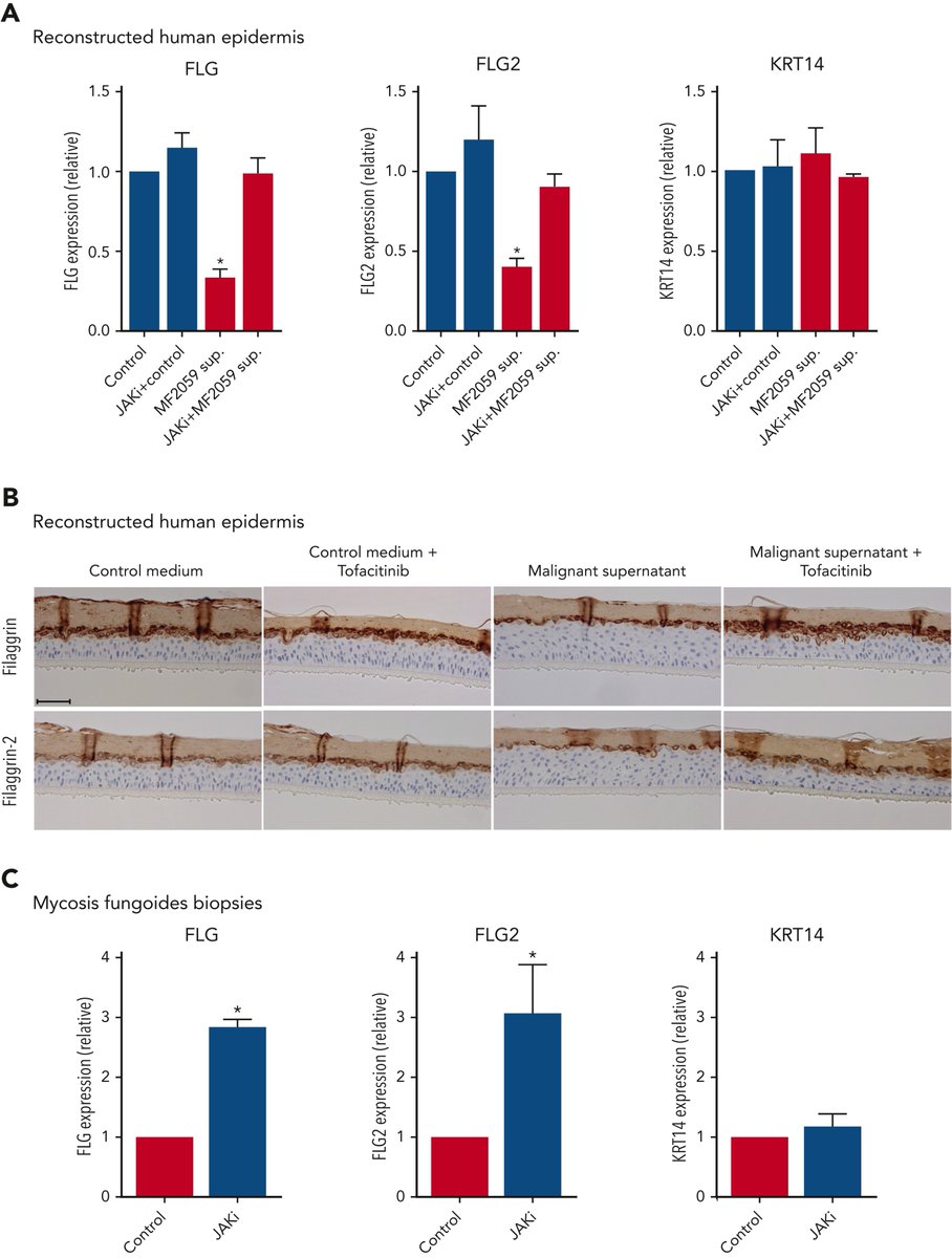
This indicates the skin barrier of CTCL patients may be improved by JAK inhibitor treatment. Indeed, treating keratinocytes, reconstructed epidermis and lesional CTCL skin biopsy explants with a clinical JAK-inhibitor (Tofacitinib), all improved FLG expression levels to normal.

This indicates the skin barrier of CTCL patients may be improved by JAK inhibitor treatment. Indeed, treating keratinocytes, reconstructed epidermis and lesional CTCL skin biopsy explants with a clinical JAK-inhibitor (Tofacitinib), all improved FLG expression levels to normal.
account_circle
Thaidakar of Ndi-Uche(@okemmin_afc) 's Twitter Profile Photo

财富 One would argue that what you refer to as dirt are actually keratinocytes that contains the melanin pigment 💁🏽‍♂️

account_circle
David Clements MD(@DrDavidClements) 's Twitter Profile Photo

Someone should do research on insulin as treatment for Xylazine wound treatment. Would migrate keratinocytes that I theorize are stagnated due to Alpha-2 agonism signal. (he says, into the universe hoping someone smarter will hear)

account_circle