ZEISS Microscopy(@zeiss_micro) 's Twitter Profileg
ZEISS Microscopy

@zeiss_micro

We are #Microscopy. All about microscopes from #ZEISS.
LinkedIn: https://t.co/4jXTmJoUMF Facebook: https://t.co/ivuXIt7d1y | Imprint & Data Privacy: https://t.co/bvddlc6GZ2

ID:46128549

linkhttps://www.zeiss.com/microscopy calendar_today10-06-2009 14:49:22

10,6K Tweets

23,4K Followers

3,8K Following

Follow People
ZEISS Microscopy(@zeiss_micro) 's Twitter Profile Photo

With the software module ZEISS Spectral RICS, we are releasing a new solution for understanding molecular dynamics of your living samples.

In addition to obtaining structural image information of your sample, unveil how proteins of interest move and interact in relation to their…

With the software module ZEISS Spectral RICS, we are releasing a new solution for understanding molecular dynamics of your living samples. In addition to obtaining structural image information of your sample, unveil how proteins of interest move and interact in relation to their…
account_circle
Dr Richard Kirby(@PlanktonPundit) 's Twitter Profile Photo

Huge spring plankton biodiversity in my net sample collected off Plymouth UK yesterday. At least: Dinoflagellata, Bacillariophyta, Haptophyta, Arthropoda, Annelida, Phoronida, Cnidaria, and Echinodermata. But what species can you see? ZEISS Microscopy

account_circle
NEMI(@ProjectNemi) 's Twitter Profile Photo

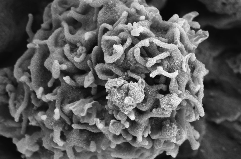
🏜️ 'Desert Rose'🌹by Ewelina Kluza from Amsterdam UMC, Amsterdam UMC

An immune cell on the endothelial surface of a mouse aorta! 🐁❤️

🔬 Imaged with a ZEISS Sigma 300 Scanning Electron Microscope ( ) ZEISS Microscopy at Amsterdam UMC

🏜️ 'Desert Rose'🌹by Ewelina Kluza from Amsterdam UMC, @amsterdamumc An immune cell on the endothelial surface of a mouse aorta! 🐁❤️ 🔬 Imaged with a ZEISS Sigma 300 Scanning Electron Microscope (#SEM) @zeiss_micro at Amsterdam UMC
account_circle
Josh Gibson, Ph.D.(@DrStrangeAnt) 's Twitter Profile Photo

MicroCT scan of a fruit fly stained with iodine, taken with Beckman Institute's brand new Zeiss Xradia ZEISS Microscopy's Xradia Versa 630 scanner! Renderings done in Amira.


account_circle
Vanderbilt Cell Imaging Shared Resource(@VUCellImaging) 's Twitter Profile Photo

Spring has sprung in Nashville! It's chilly, but the flowers are blooming, and everything is green. We found this 'helicopter seed' on campus and couldn't resist checking its autofluorescence for . ZEISS Microscopy confocal, 20x/0.8. Max. Intensity Projection.

Spring has sprung in Nashville! It's chilly, but the flowers are blooming, and everything is green. We found this 'helicopter seed' on campus and couldn't resist checking its autofluorescence for #FluorescenceFriday. @zeiss_micro confocal, 20x/0.8. Max. Intensity Projection.
account_circle
Danielle Beckman(@DaniBeckman) 's Twitter Profile Photo

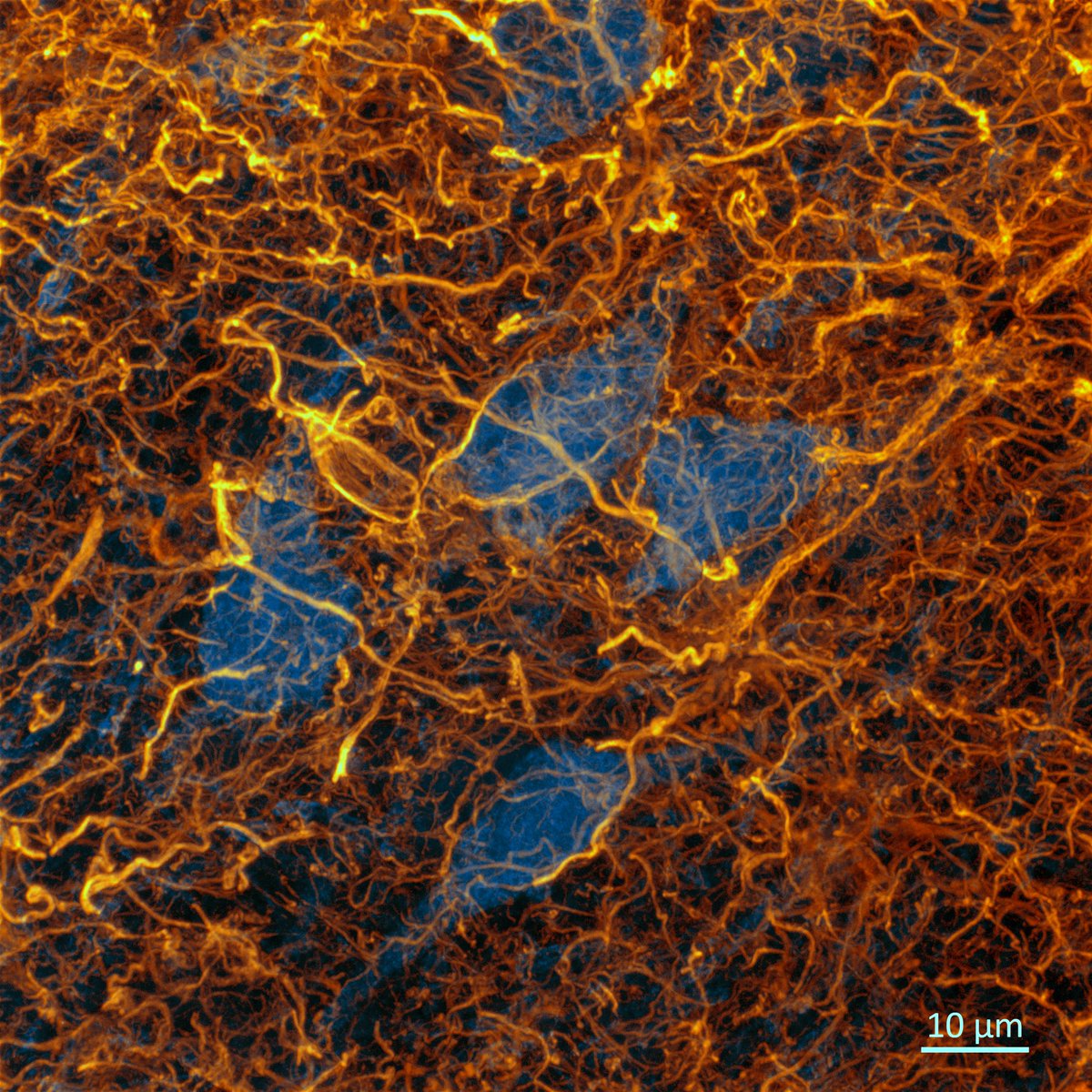
Astrogliosis (🟡) in the human brain 🧠🔬😨. One of the processes occurring in neuroinflammation. These neurons (🔵) are condemned to die...
Image taken from the frontal cortex of a patient with advanced Alzheimer's disease (Braak Stage VI)
ZEISS Microscopy

Astrogliosis (🟡) in the human brain 🧠🔬😨. One of the processes occurring in neuroinflammation. These neurons (🔵) are condemned to die... Image taken from the frontal cortex of a patient with advanced Alzheimer's disease (Braak Stage VI) @zeiss_micro #neuroinflammation
account_circle
EXCITE(@EXCITE_network) 's Twitter Profile Photo

Happy ! map of a synthetic lithium slag showing crystal orientation.

🔬 Imaged with a ZEISS Gemini 450 (ZEISS Microscopy) at Utrecht University (@UniUtrecht) by Maartje Hamers for EXCITE TNA user Cindytami Rachmawati, TU Bergakademie Freiberg
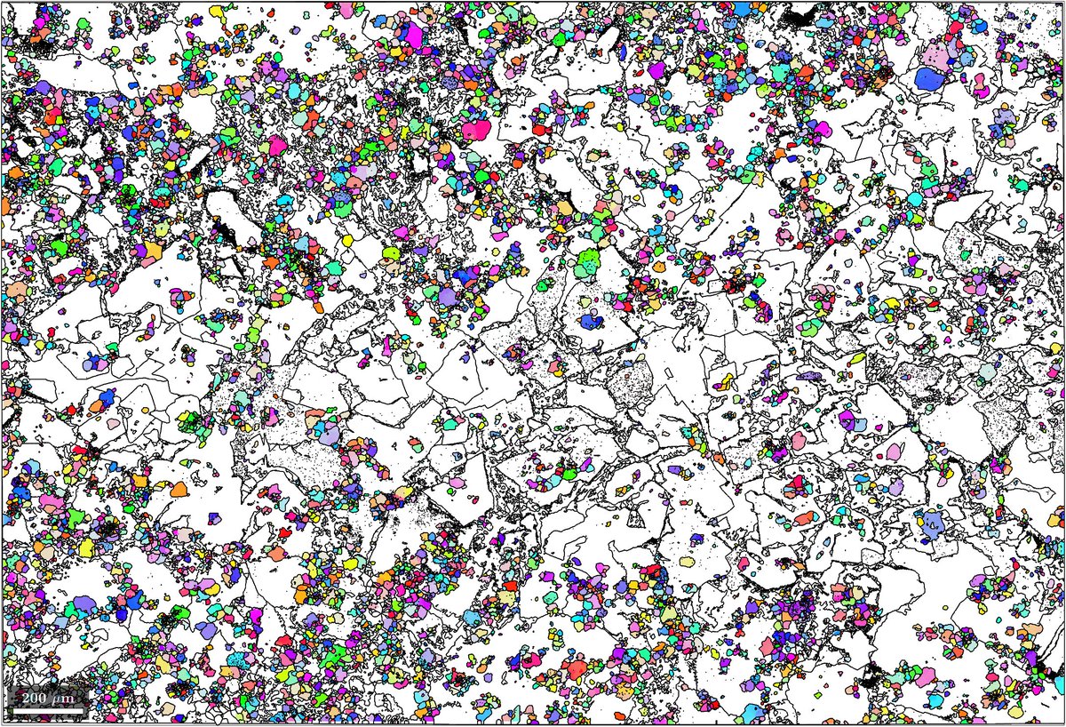
Happy #ThinSectionThursday! #EBSD map of a synthetic lithium slag showing #spinel crystal orientation. 🔬 Imaged with a ZEISS Gemini 450 #SEM (@zeiss_micro) at Utrecht University (@UniUtrecht) by Maartje Hamers for @EXCITE_network TNA user Cindytami Rachmawati, @TUBergakademie
account_circle
Rosario in Paris ᥫ᭡(@chayito09) 's Twitter Profile Photo

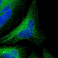
Estas células COS-7 (células de fibroblasto derivadas del tejido renal del mono verde africano) fueron teñidas con fluoróforos diferentes para capturar el complejo mundo de la biología.
🧫🔬

©️ ZEISS Microscopy
account_circle
micROCKScopica(@micROCKScopica) 's Twitter Profile Photo

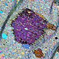
Playing with the hi-res scans of an epidote blueschist from Syros, Greece.
I don't know what is better, epidote or glaucophane?
Thanks ZEISS Microscopy for making this possible.

Playing with the hi-res scans of an epidote blueschist from Syros, Greece. I don't know what is better, epidote or glaucophane? Thanks @zeiss_micro for making this possible.
account_circle
NEMI(@ProjectNemi) 's Twitter Profile Photo

📆 April image of the month: 'The Ties That Bind Us' by Irene Schimmel, Amsterdam UMC (Amsterdam UMC).

🫀Extracellular matrix in a mouse heart.

🔬Imaged with a ZEISS Gemini Sigma 300 (@zeiss_micro) at the Electron Microscopy Center Amsterdam (EMCA), Amsterdam UMC

📆 April image of the month: 'The Ties That Bind Us' by Irene Schimmel, Amsterdam UMC (@amsterdamumc). 🫀Extracellular matrix in a mouse heart. 🔬Imaged with a ZEISS Gemini Sigma 300 #SEM (@zeiss_micro) at the Electron Microscopy Center Amsterdam (EMCA), Amsterdam UMC
account_circle
Danielle Beckman(@DaniBeckman) 's Twitter Profile Photo

It's !
It is always a good Friday with some microglia! 🧠🤓
In this photo, these immune cells of the brain (🟡) are reacting to some neurons (🔵) accumulating p-tau (🔴) in an monkey brain.
with ZEISS Microscopy

It's #FluorescenceFriday! It is always a good Friday with some microglia! 🧠🤓 In this photo, these immune cells of the brain (🟡) are reacting to some neurons (🔵) accumulating p-tau (🔴) in an #Alzheimer monkey brain. with @zeiss_micro
account_circle
Aberdeen Microscopy(@Abdn_Micro) 's Twitter Profile Photo

Delighted to have our new ZEISS Microscopy Lightsheet 7 delivered earlier this month! Thanks to a successful Biotechnology and Biological Sciences Research grant and a lot of hard work we’ve finally been able to bring this technology to . If you’d like to know how it could enhance your , get in touch 🙂

Delighted to have our new @zeiss_micro Lightsheet 7 delivered earlier this month! Thanks to a successful @BBSRC grant and a lot of hard work we’ve finally been able to bring this technology to #Aberdeen. If you’d like to know how it could enhance your #research, get in touch 🙂
account_circle
ZEISS Microscopy(@zeiss_micro) 's Twitter Profile Photo

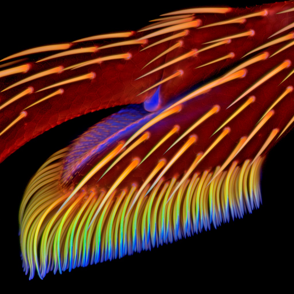
Spring is coming! 🐞🔬
Using confocal laser scanning microscopy and atomic force microscopy to investigate the legs of a ladybird beetle (Coccinella septempunctata)

© Dr. Jan Michels, Institute of Zoology, Christian-Albrechts-Universität zu Kiel

Spring is coming! 🐞🔬 Using confocal laser scanning microscopy and atomic force microscopy to investigate the legs of a ladybird beetle (Coccinella septempunctata) © Dr. Jan Michels, Institute of Zoology, Christian-Albrechts-Universität zu Kiel #ladybird #microscopy #spring
account_circle
Danielle Beckman(@DaniBeckman) 's Twitter Profile Photo

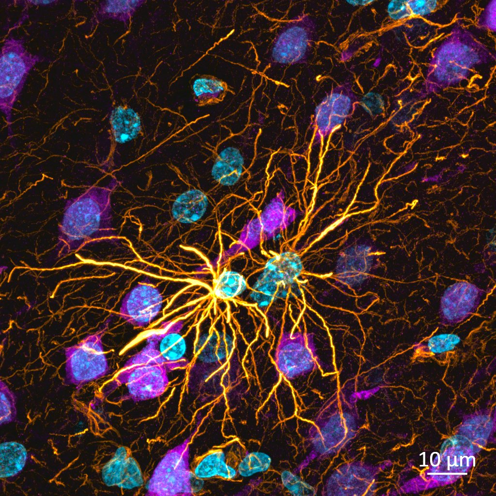
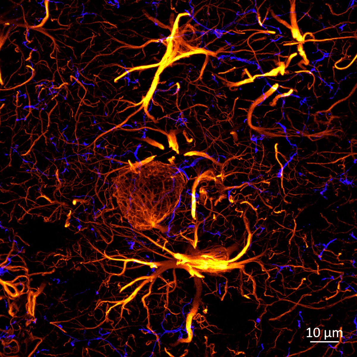
Oof, what these astrocytes are doing? They stole the show in this case; microglia are barely appearing
🔵microglia (iba1)🟡astrocytes (gfap) ZEISS Microscopy

Oof, what these astrocytes are doing? They stole the show in this case; microglia are barely appearing 🔵microglia (iba1)🟡astrocytes (gfap) #neuroscience @zeiss_micro
account_circle