Hao Yin(@HaoYin20) 's Twitter Profile Photo

Amazing😎Hematoxylin/Eosin-Mimetic NonDestructive 3D Histology of FFPE archived tissue

TOPRO3+Eosin➡️
Optical clearing with ethyl cinnamate➡️
Light-sheet microscopy➡️
Converted to HE-like image with FalseColor-Python➡️
Bioimage analysis with Napari napari🔬

What's the

account_circle
Brant Bassam(@BrantBassamPhD) 's Twitter Profile Photo

We’re all wondering what the future of (ST) has in store. Well, I think the near future will look a lot like the new 6000-plex dataset reviewed here (link to the original source at the end of this post).

SAMPLE
An entire 100 mm² intact 5 µm FFPE section
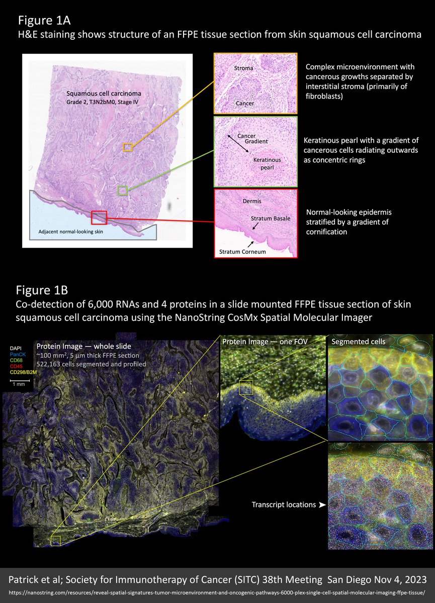
We’re all wondering what the future of #spatialtranscriptomics (ST) has in store. Well, I think the near future will look a lot like the new 6000-plex  dataset reviewed here (link to the original source at the end of this post).

SAMPLE
An entire 100 mm² intact 5 µm FFPE section
account_circle
10x Genomics(@10xGenomics) 's Twitter Profile Photo

3 reasons why we think CytAssist can take your spatial studies further:

1. Simplified sample processing from glass slides, including pre-sectioned or archived FFPE tissue
2. Robust, consistent spatial barcoding for high-quality data
3. 👀

bit.ly/3wFXXKB

account_circle
🧬株式会社CyberomiX🧬|ゲノミクスで未来を変えるカンパニー(@CyberomiX) 's Twitter Profile Photo


✨さいぼNEWS!〖 速報 〗 🙌✨


みなさま〜!!
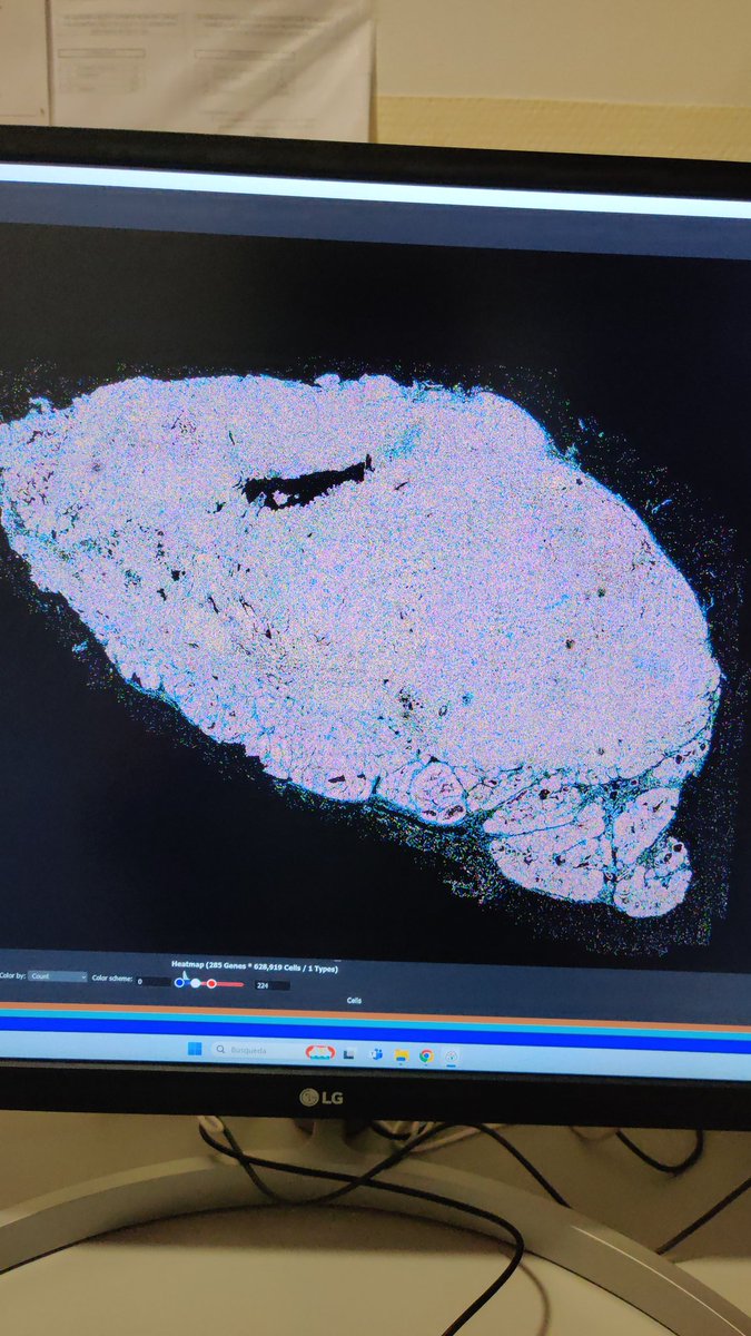
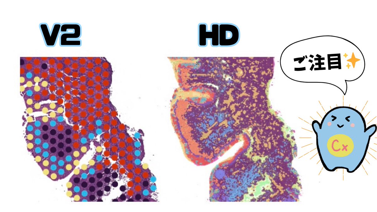
この度、高解像度の新プラットフォームVisium HD初のデータ解析が終わりました👏㊗️🎉

ぜひ‼️
下記の画像をご覧ください🧑‍🏫✨
旧プラットフォームVisium FFPE v2との違いは一目瞭然です👀💫

気になる方はいますぐ🧬株式会社CyberomiX🧬|ゲノミクスで未来を変えるカンパニー にご連絡を📢 ˎˊ˗
/
✨さいぼNEWS!〖 速報 〗 🙌✨
\

みなさま〜!!
この度、高解像度の新プラットフォームVisium HD初のデータ解析が終わりました👏㊗️🎉

ぜひ‼️
下記の画像をご覧ください🧑‍🏫✨
旧プラットフォームVisium FFPE v2との違いは一目瞭然です👀💫

気になる方はいますぐ@CyberomiX にご連絡を📢 ˎˊ˗
account_circle
Hao Yin(@HaoYin20) 's Twitter Profile Photo

Patho-DBiT
Spatial on FFPE section👹

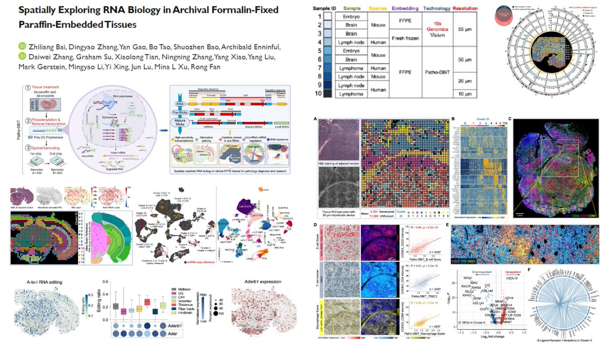
Now tissues can be mapped by RNA variants or non-coding RNAs🤯

Whole transcriptome sequencing covers
mRNA
LincRNA
microRNA
sn/snoRNA
105 events/85 genes/spot🤓

RNA

Patho-DBiT
Spatial #RNAWorld on FFPE section👹

Now tissues can be mapped by RNA variants or non-coding RNAs🤯

Whole transcriptome sequencing covers 
mRNA
LincRNA
microRNA
sn/snoRNA
#AlternativeSplicing 105 events/85 genes/spot🤓
#RNAEditing
RNA #SingleNucleotideVariation
account_circle
Cytopathology(@CytopathologyJ) 's Twitter Profile Photo

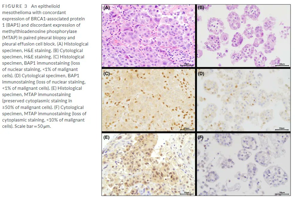
In this study multiple IHC markers in 59 pleural were compared in paired specimens. Biopsies and FFPE pleural effusion cell blocks were concordant in most cases.
buff.ly/3PGb19X

In this #OpenAccess study multiple IHC markers in 59 pleural #mesotheliomas were compared in paired specimens. Biopsies and FFPE pleural effusion cell blocks were concordant in most cases. #pulmpath #CytoHistoCorrelation #CytoJ
buff.ly/3PGb19X
account_circle
Akoya Biosciences(@AkoyaBio) 's Twitter Profile Photo

Today's features a whole slide of a human FFPE tumor labeled with 100+ antibodies. Imaged on the system, it reveals 14 distinct cell types and four unique tumor regions.

Inspired by @Aruthak's award-winning , the

Today's #TissueTuesday features a whole slide of a human FFPE #headandneck tumor labeled with 100+ antibodies. Imaged on the #PhenoCyclerFusion system, it reveals 14 distinct cell types and four unique tumor regions. 

Inspired by @Aruthak's award-winning #cancerresearch, the
account_circle
ClaretBio(@ClaretBio) 's Twitter Profile Photo

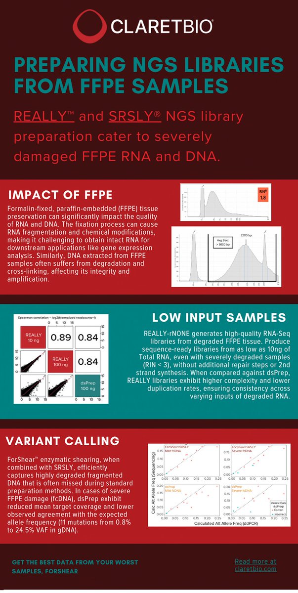
At Claret, we get it - sequencing isn't always a walk in the park. When it comes to library prepping FFPE samples, we know the struggle is real.
Discover how Claret's NGS kits perform with FFPE RNA and DNA samples.

At Claret, we get it - sequencing isn't always a walk in the park. When it comes to library prepping FFPE samples, we know the struggle is real. 
Discover how Claret's NGS kits perform with FFPE RNA and DNA samples.
#SRSLY #REALLY #FFPE #libraryprep #NGS
account_circle
Akoya Biosciences(@AkoyaBio) 's Twitter Profile Photo

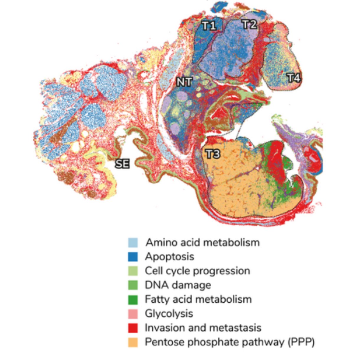
Today, we're featuring a figure that shows whole-slide of a human FFPE squamous cell carcinoma with an ultrahigh-plex antibody panel using our solution.

It reveals four distinct tumor regions with varying metabolic and

Today, we're featuring a figure that shows whole-slide #singlecell #spatialphenotyping of a human FFPE #headandneck squamous cell carcinoma with an ultrahigh-plex antibody panel using our #spatialbiology solution.

It reveals four distinct tumor regions with varying metabolic and
account_circle
Rong Fan(@RongFan8) 's Twitter Profile Photo

Super-resolved single-cell atlas of a human lymphoma FFPE tissue slide profiled using our latest Patho-DBiT technology and the novel pipeline iStar developed by Mingyao Li . Just too psyched to not share 🤩🤩
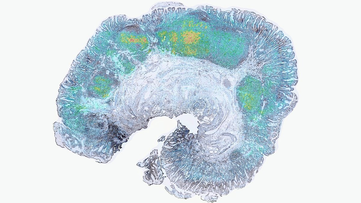
Super-resolved single-cell atlas of a human lymphoma FFPE tissue slide profiled using our latest Patho-DBiT technology and the novel pipeline iStar developed by @DrMingyaoLi . Just too psyched to not share 🤩🤩
account_circle
Rong Fan(@RongFan8) 's Twitter Profile Photo

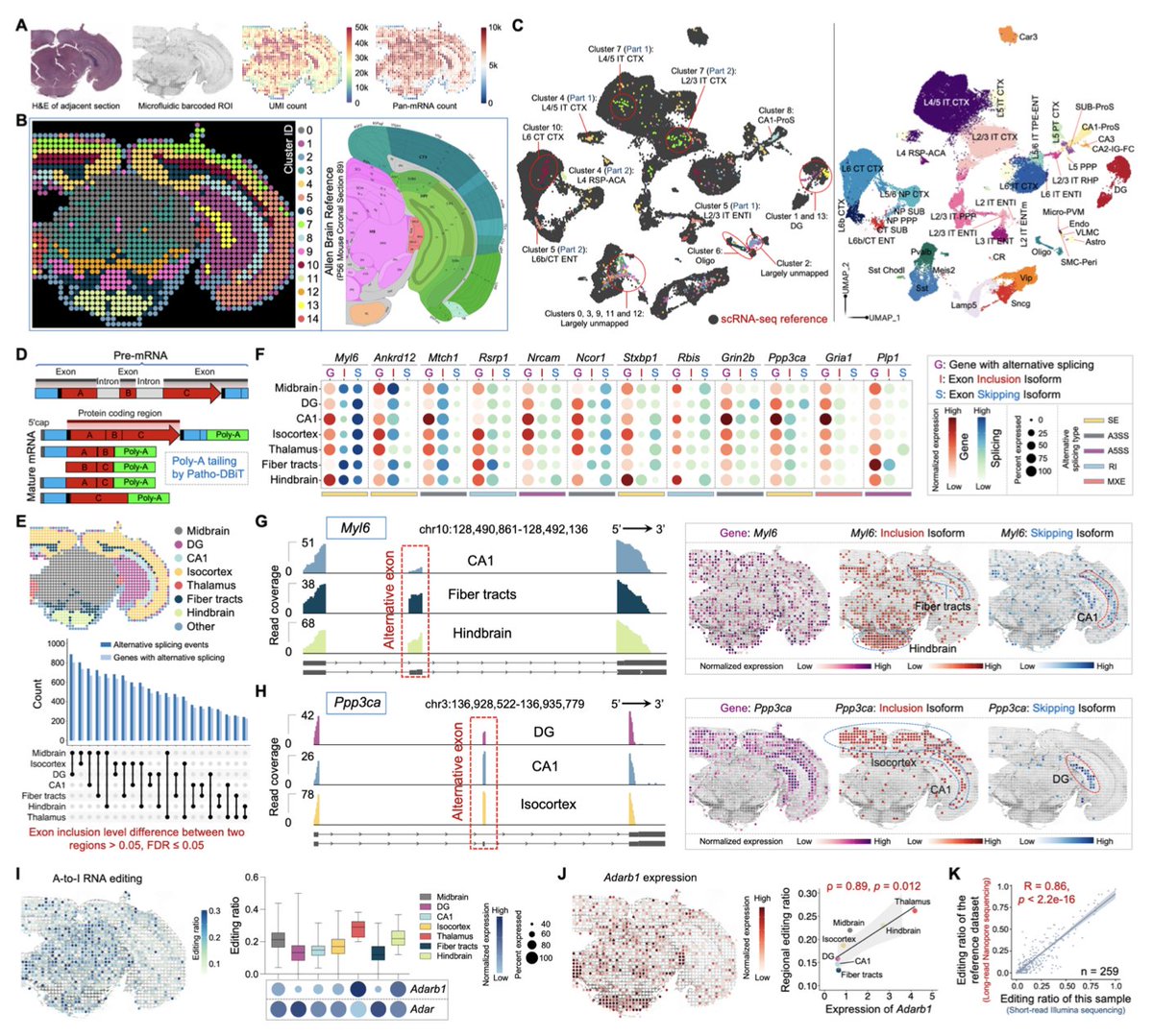
we mapped alternative splicing over the entire hemisphere of mouse brain (FFPE) and discovered a lot of region-specific isoforms :)

we mapped alternative splicing over the entire hemisphere of mouse brain (FFPE) and discovered a lot of region-specific isoforms :)
account_circle
Akoya Biosciences(@AkoyaBio) 's Twitter Profile Photo

Wishing you a filled with striking red roses and heart-shaped tissues! 🌹

🌹The roses are made from FFPE breast tissue with a pattern of red and green stains.

💗The heart-shaped tissues are made from FFPE breast cancer using PhenoCycler-Fusion.

Conrad Lee
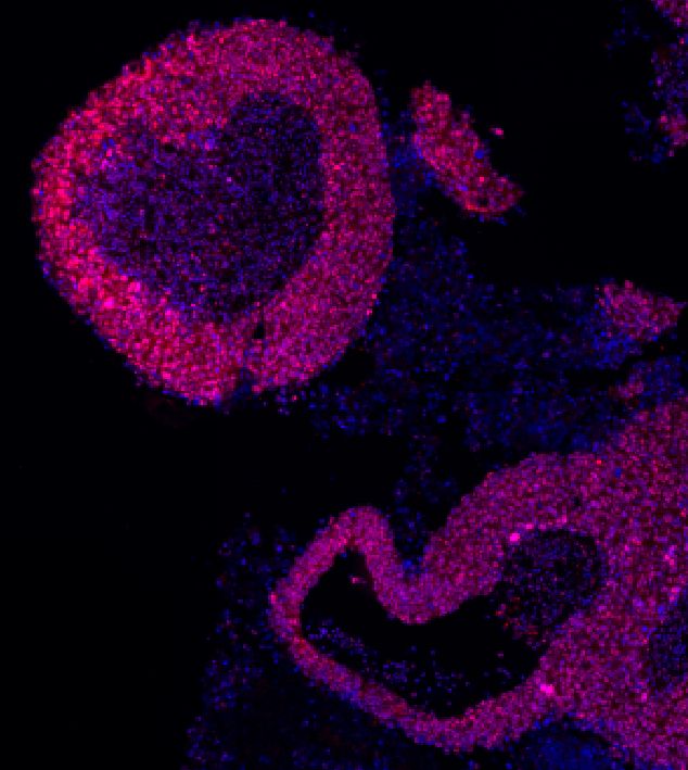
Wishing you a #ValentinesDay filled with striking red roses and heart-shaped tissues!  🌹 

🌹The roses are made from FFPE breast tissue with a pattern of red and green stains.  

💗The heart-shaped tissues are made from FFPE breast cancer using PhenoCycler-Fusion.

@conrad45605
account_circle
Vesiculab Ltd(@Vesiculab) 's Twitter Profile Photo

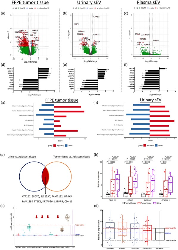
Small EVs as biomarkers for bladder cancer: Ken Pienta at JHU et al conducted RNA sequencing for FFPE tumor tissues and small EVs from matched tissue explants, urine, and plasma in BCa patients isevjournals.onlinelibrary.wiley.com/doi/full/10.10…

Small EVs as biomarkers for bladder cancer: Ken Pienta at JHU et al conducted RNA sequencing for FFPE tumor tissues and small EVs from matched tissue explants, urine, and plasma in BCa patients isevjournals.onlinelibrary.wiley.com/doi/full/10.10… #extracellularvesicles #exosomes #rna #liquidbiopsy #Vesiculab
account_circle
Front Line Genomics(@FLGenomics) 's Twitter Profile Photo

Interested in automated sample prep for from FFPE samples? Nathan Pereira, Product Manager at S2 Genomics will be presenting the new Singulator 200+ on stage at , which automates the whole process. Register now: hubs.la/Q02y5zQq0
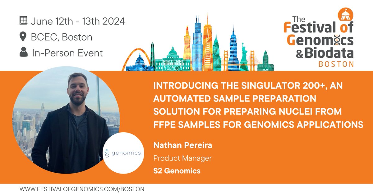
Interested in automated sample prep for #singlenuclei from FFPE samples? Nathan Pereira, Product Manager at @S2Genomics will be presenting the new Singulator 200+ on stage at #FOGBoston, which automates the whole process. Register now: hubs.la/Q02y5zQq0
account_circle
Akoya Biosciences(@AkoyaBio) 's Twitter Profile Photo

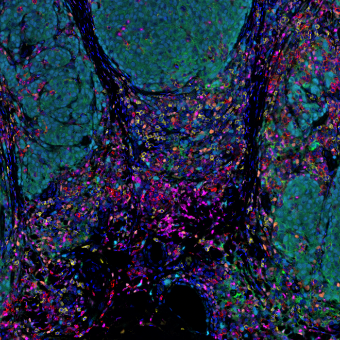
For today's , we're sharing a human melanoma FFPE tissue stained w/ a 6-plex MOTiF PD-1/PD-L1 panel auto kit & imaged on .

Learn about our kits crucial for melanoma samples in translational research. bit.ly/3wdqHuc

For today's #TissueTuesday, we're sharing a human melanoma FFPE tissue stained w/ a 6-plex MOTiF PD-1/PD-L1 panel auto #melanoma kit & imaged on #PhenoImagerHT.

Learn about our kits crucial for melanoma samples in translational #immunooncology research. bit.ly/3wdqHuc
account_circle
Akoya Biosciences(@AkoyaBio) 's Twitter Profile Photo

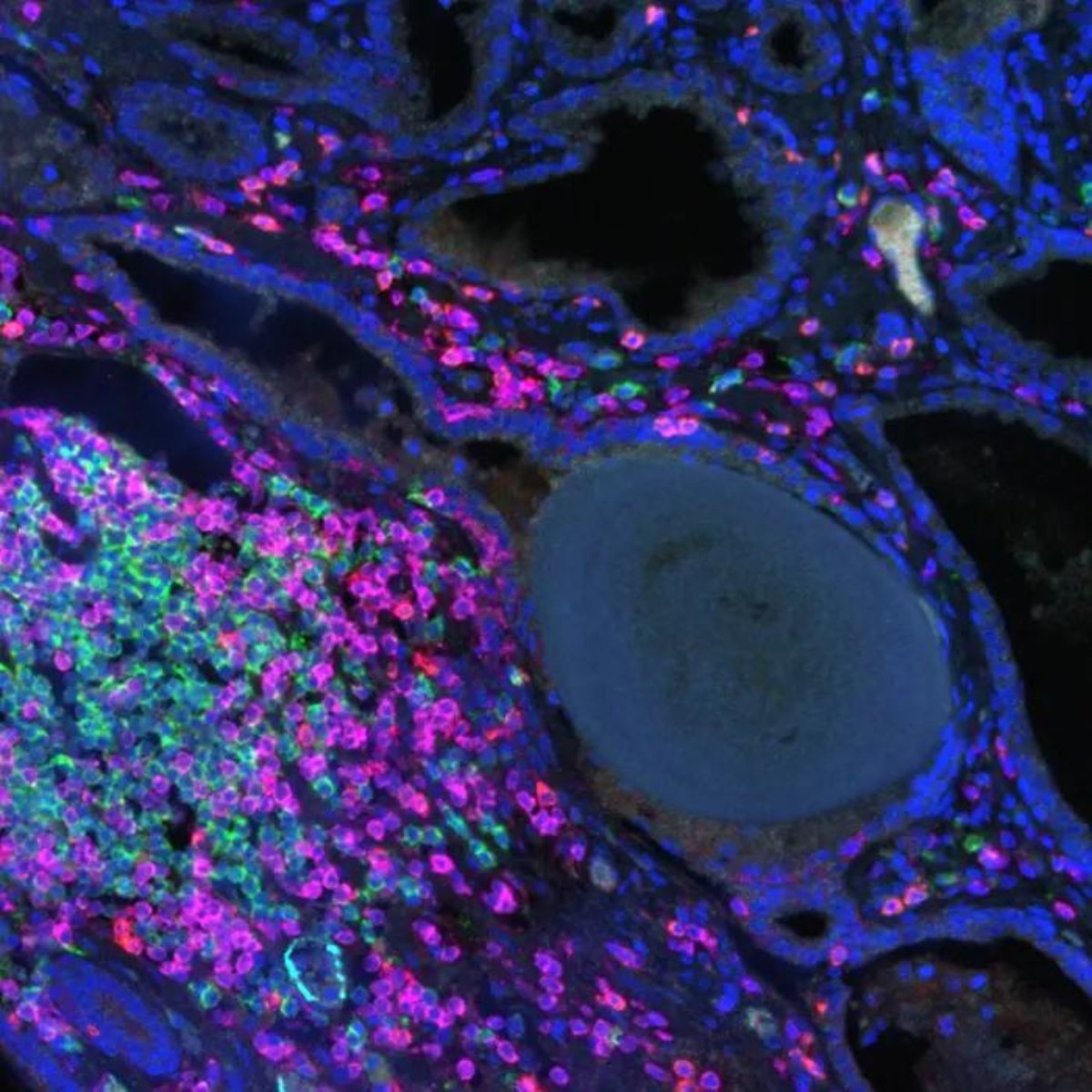
Today's showcases FFPE tissue stained with an Opal 6-plex panel (CD4, CD8, CD19, CD21, DC-LAMP, and PNAd), revealing a Type V (TLS).

Thank you, Jessica Duarte, for using our solution to generate this

Today's #TissueTuesday showcases #prostatecancer FFPE tissue stained with an Opal 6-plex panel (CD4, CD8, CD19, CD21, DC-LAMP, and PNAd), revealing a Type V #tertiarylymphoidstructures (TLS).

Thank you, @JessDGDuarte, for using our #PhenoImager solution to generate this
account_circle