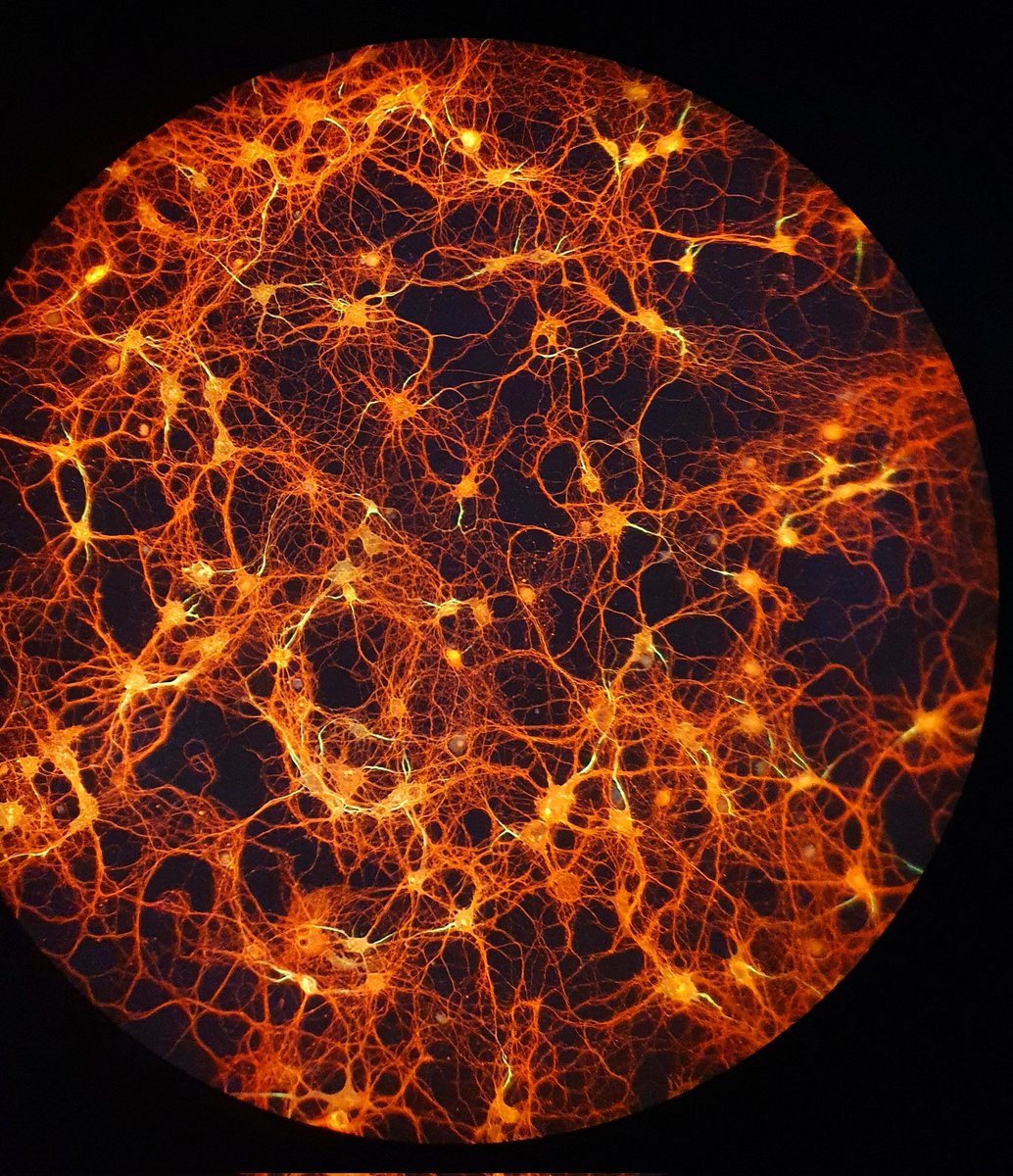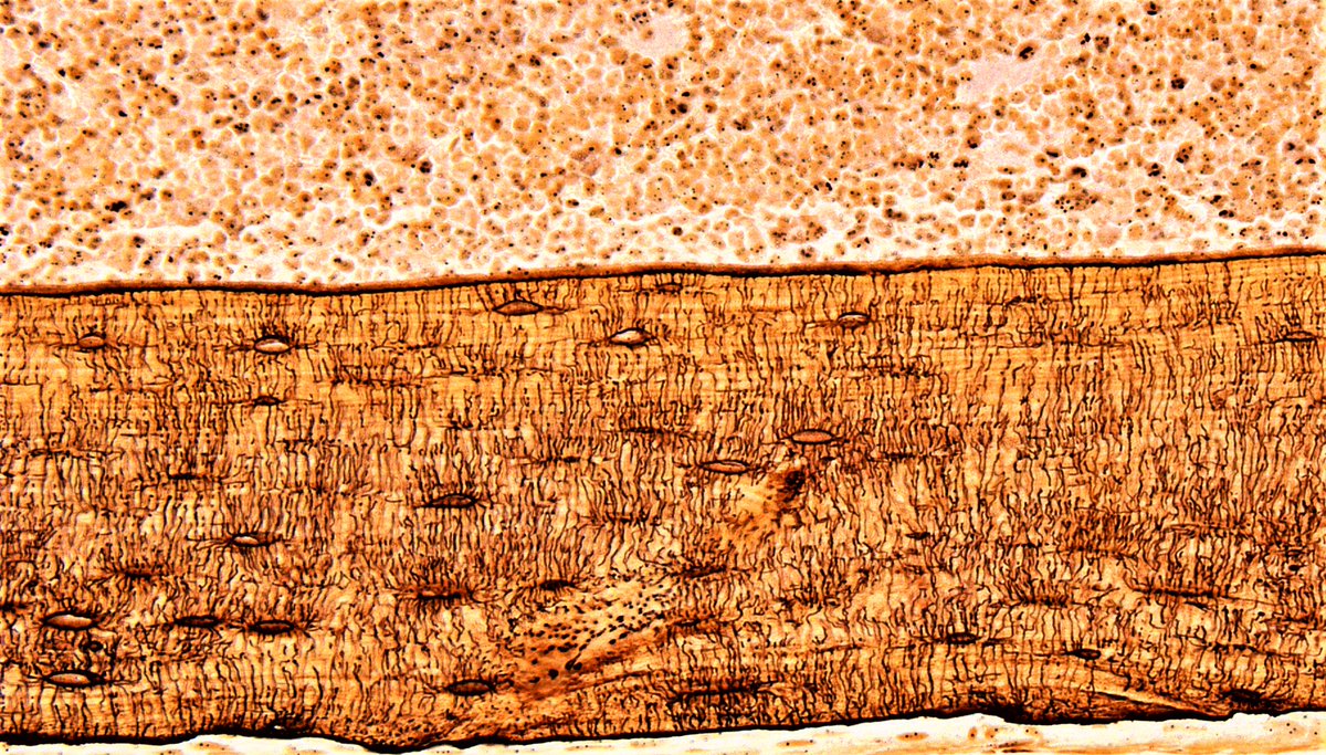
A light-sheet Z-stack tour through a 6-month-old female 5xFAD mouse hemibrain. Bright spots are Aβ plaques stained using Congo red 🔬 #MicroscopyMonday


Found a little demo I wrote for microscope simulation during the time I was learning microscopy, still fun to play with. #WolframDemo #MicroscopyMonday


Creating my first 3D model of my optical setup in blender, following the fantastic tutorial by Ryo Mizuta Graphics
#MicroscopyMonday
Can't decide whether it is #MicroscopyMonday , #MovieMonday , or #Migration Monday? Why not have it all? Here's a movie of Dictyostelium (Dicty) with fluorescently labeled nuclei migrating through microfluidic devices with small constrictions (~2x5um^2). Credit: Jacob Odell

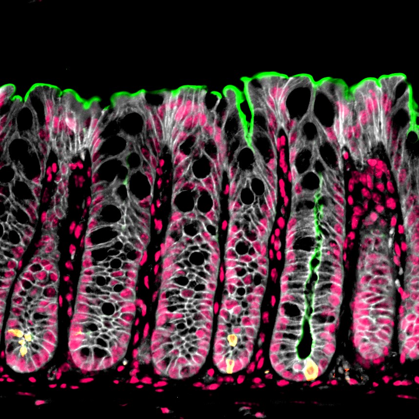
Happy #MicroscopyMonday ! Immunofluorescence staining of the colon highlighting the brush border (green), cell membrane (white), tuft cells (yellow), and nuclei (pink).
#microscopy #sciart #pathart #bioart #histoart #intestine

#MicroscopyMonday
Others: Let’s take some beautiful 4D multi-colour images!
Me at the microscope:

One more video series celebrating #MicroscopyMonday 🙂. To give you an impression of the raw data we show you the transition of cytoskeleton targets to synaptic targets and markers, with a zoom-in into an individual synapse, starting with the cytoskeleton in a ~10 um^2 FoV 1/4

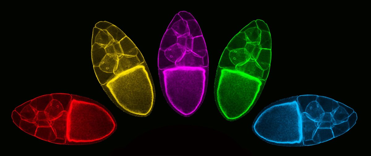
It's a rainy day St. Jude Research in Memphis, so here is a science rainbow to chase the clouds away. This is a #SciArt piece by former postdoc Suresh Marada. Image shows a Drosophila egg chamber pseudocolored using photoshop. Happy #MicroscopyMonday !

Just under the wire for #MicroscopyMonday , here are mouse photoreceptors labeled with transducin antibody. Does someone know what the small brighter spots are, near the connecting cilia (examples with yellow arrowheads)?


I'm thrilled to have won the SLB Image Contest 🏆, in celebration of the #InternationalDayofImmunology 2024 featuring Hypereosinophilia at High Resolution! Society for Leukocyte Biology #WorldImmunologyDay #immunology
#MicroscopyMonday


21 day old HCO cultures stained for E-CAD (green) CD34 (red) and counterstained for DAPI (blue). #microscopymonday


This weekend I found this massive fibroblasts infected with 16 Trypanosoma cruzi parasites. Right on time for #MicroscopyMonday BCM_TropMed


Happy #MicroscopyMonday !
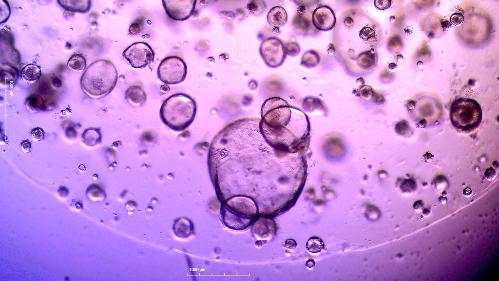
Enjoy these swell mouse organoids during a rotavirus infection
#organoids #microscopy

A big hello to science twitter from the new Brandeis Microscopy feed. We will be sharing all things to do with the light and electron microscopy facilities on this page. Follow to hear more about the exciting science being done at our facilities #microscopymonday


Get ready to level up your live cell actin imaging game with SPY555-FastAct! 🤩🔬
Fast turnover F-actin detection, staining performed in 2h, high brightness and photostability!
Check it out now 👉 spirochrome.com/product/spy555…
#cellimaging #microscopy #SPYprobes #MicroscopyMonday
