Jacques Morcos MD FRCS FAANS
@jacquesmorcosmd
Professor & Chairman of #Neurosurgery, Director #Cerebrovascular #SkullBaseSurgery, University of Texas Health Sciences Houston @UTHealthHouston @UTHNeuro
ID:1265484179489411074
https://med.uth.edu/neurosciences/jacques-j-morcos-md-frcs-faans/ 27-05-2020 03:25:34
1,2K Tweets
8,1K Followers
94 Following
Follow People

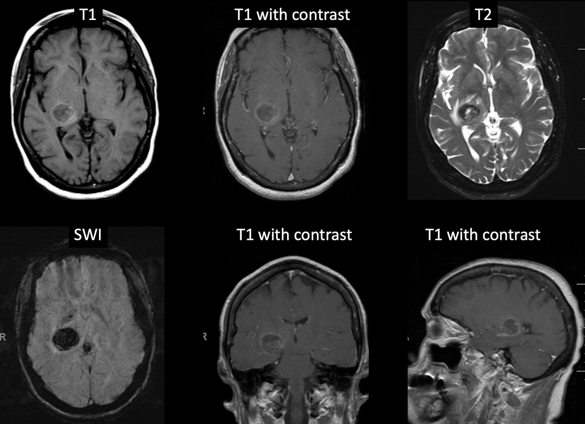
#MorcosChallenge MRI at 6 weeks showed resolving area of hemorrhage. Left hemiparesis and hemianesthesia resolved but she continued to have left homonymous inferior quadrantopsia Eva Wu UTHealthNeurosciences Neurosurgical Atlas Medical Clinical Case BNTA CV Section @neuroangioA @neurosurgA

#MorcosChallenge MRI w/ lesion in R pulvinar extending into posterior limb of internal capsule w/ intrinsic areas of high T1 signal&SWI artifact consistent w/ blood products. No enhancement to suggest underlying mass. Plan to repeat MRI in 4-6wks Eva Wu UTHealthNeurosciences CV Section

#MorcosChallenge What does the MRI show? What is the suspected dx? How would you manage pt? Eva Wu UTHealthNeurosciences Neurosurgical Atlas @neuroangioA @neurosurgA Neuroangio BNTA Medical Clinical Case CV Section @nasbsorg @neuronotes @nansig1 NeurExplain @uncleharveynsg @neurosurgeryspr

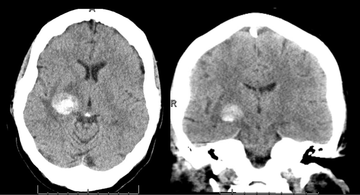
#MorcosChallenge CTH w/ hyperdensity&surrounding hypodensity in pulvinar of R thalamus w/ extension into posterior limb of the internal capsule. Given h/o of HTN&SBP180s on arrival, likely represents hypertensive ICH. MRI brain obtained to r/o underlying lesion Eva Wu UTHealthNeurosciences

#MorcosChallenge 67F w/ h/o HTN, presented to ED w/ SBP180s, left homonymous inferior quandrantopsia, left hemiparesis&hemianesthesia. What does the CT show? What do you think the ddx is? What would you do next? Eva Wu UTHealthNeurosciences Medical Clinical Case CV Section BNTA @neuroangioA


…with the help of the UTHealth Houston UT Neurosurgery team!!! Thank you Nitin Tandon ! A wonderful year ahead. See you all in Boston, April 25-28, AANS 2025! AANS

Thank you Angela M Richardson, MD, PhD ! I had a lot of fun talking to the #E2M early to mid career neurosurgeons today at #AANS2024 and honored to be the guest to that luncheon. Always fun to reflect on the moral underpinnings of being a neurosurgeon, along with the value of #mentorship

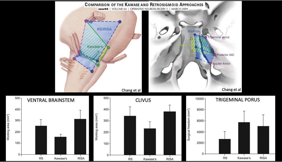
#MorcosChallenge Lesions involving trigeminal porus or Meckel’s cave can be access thru Kawase or RISA. Kawase if lesion predominantly in mfossa w/ little pfossa component. RISA if lesion predominantly in pfossa w/ little mfossa component.Retrosig if lesion limited to pfossa only

I look forward to be teaching this great hands-on course AANS in Chicago! We do not pay enough attention in general to #microsurgery subtleties and #ergonomics . Badly needed. Come join us.

#MorcosChallenge When should Kawase vs Retrosig vs RISA be used? Eva Wu UTHealthNeurosciences @nasbsorg Neurosurgical Atlas @neuronotes Medical Clinical Case BNTA @nansig1 NeurExplain @uncleharveynsg @neurosurgeryspr @nwebinar @sns_neurosurg Brainbook @basicneurosurge @neurosurgerynch


#MorcosChallenge What is the difference in exposure with Kawase vs retrosig vs RISA? Eva Wu UTHealthNeurosciences @nasbsorg Neurosurgical Atlas @neuronotes Medical Clinical Case BNTA @nansig1 NeurExplain @uncleharveynsg @neurosurgeryspr @nwebinar @sns_neurosurg Brainbook @basicneurosurge

#MorcosChallenge The bone drilled in the Kawase from above and Kawase from below (RISA) are the same. The petrous apex is removed in both and in both approaches,you work between CN5 and 7/8 Eva Wu UTHealthNeurosciences @nasbsorg Neurosurgical Atlas @neuronotes Medical Clinical Case BNTA @nansig1
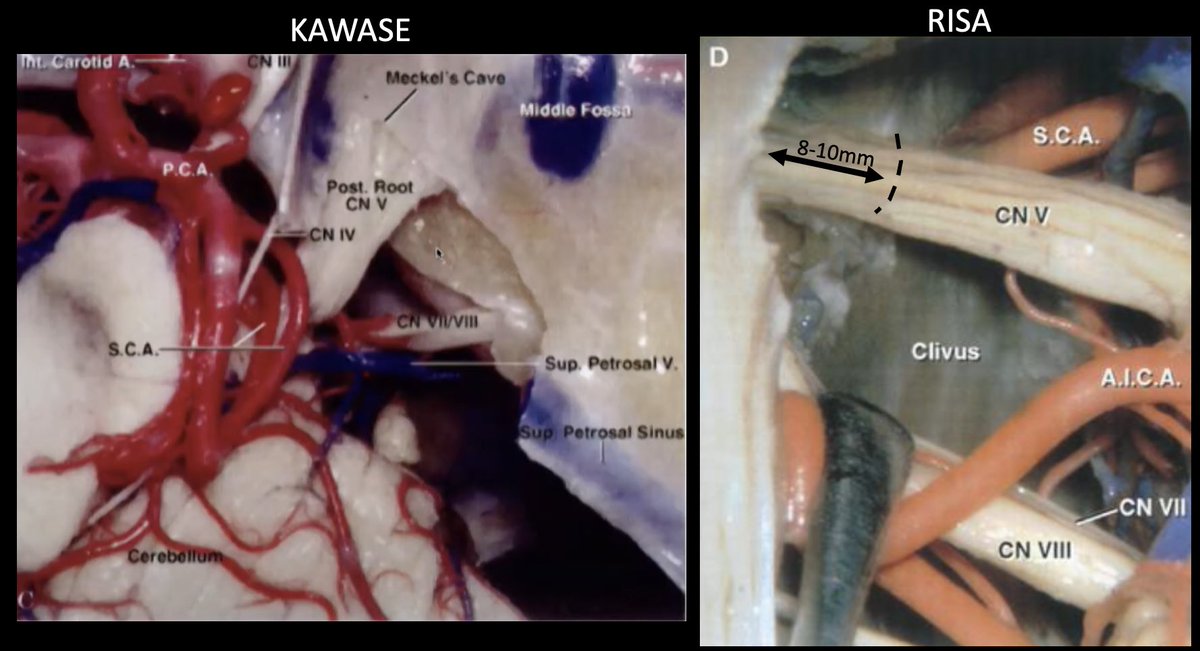

#MorcosChallenge How does the bone drilled in the RISA differ from the bone drilled in the Kawase? Eva Wu UTHealthNeurosciences Neurosurgical Atlas Young Neurosurgeons @neurosurgery101 @neurosurgA @brainschool101 @neuroanatcollab Medical Clinical Case BNTA @neuronotes Brainbook @basicneurosurge


#MorcosChallenge Drilling the suprameatal tubercle allows additional 8-10mm view of CN5 within Meckel’s cave improving access to mfossa & surgical freedom around porus of CN5. Does not increase exposure of ventral brainstem/clivus Eva Wu UTHealthNeurosciences Medical Clinical Case @nasbsorg
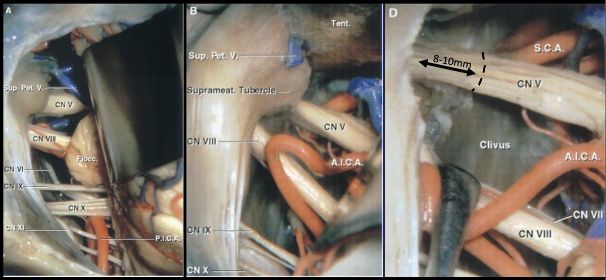

#MorcosChallenge What additional exposure does drilling the suprameatal tubercle in RISA get you? Eva Wu UTHealthNeurosciences Neurosurgical Atlas Medical Clinical Case @neuronotes Brainbook @basicneurosurge @nansig1 @sns_neurosurg @dandywedns @neurosurgerynch @neurosurgeryspr NeurExplain

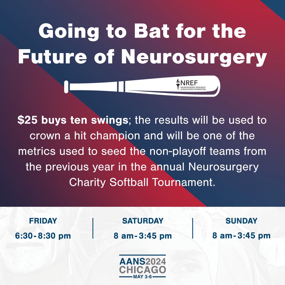
Make sure to visit the NREF exhibit hall to participate in the “Going to Bat for the Future of Neurosurgery” at the #AANS2024 conference! AANS Isaac Yang MD UCLA Neurosurgery 🧠 UCLA Health Katie O'Meara Orrico Jacques Morcos MD FRCS FAANS #whatmatters2me
