Jing Liu, MD
@JingLiu_MD
Interventional Cardiologist @BCMHeart | Houston VA | IM alum @BCM_InternalMed | #WIC
ID:1118327876103884800
17-04-2019 01:38:37
236 Tweets
267 Followers
128 Following

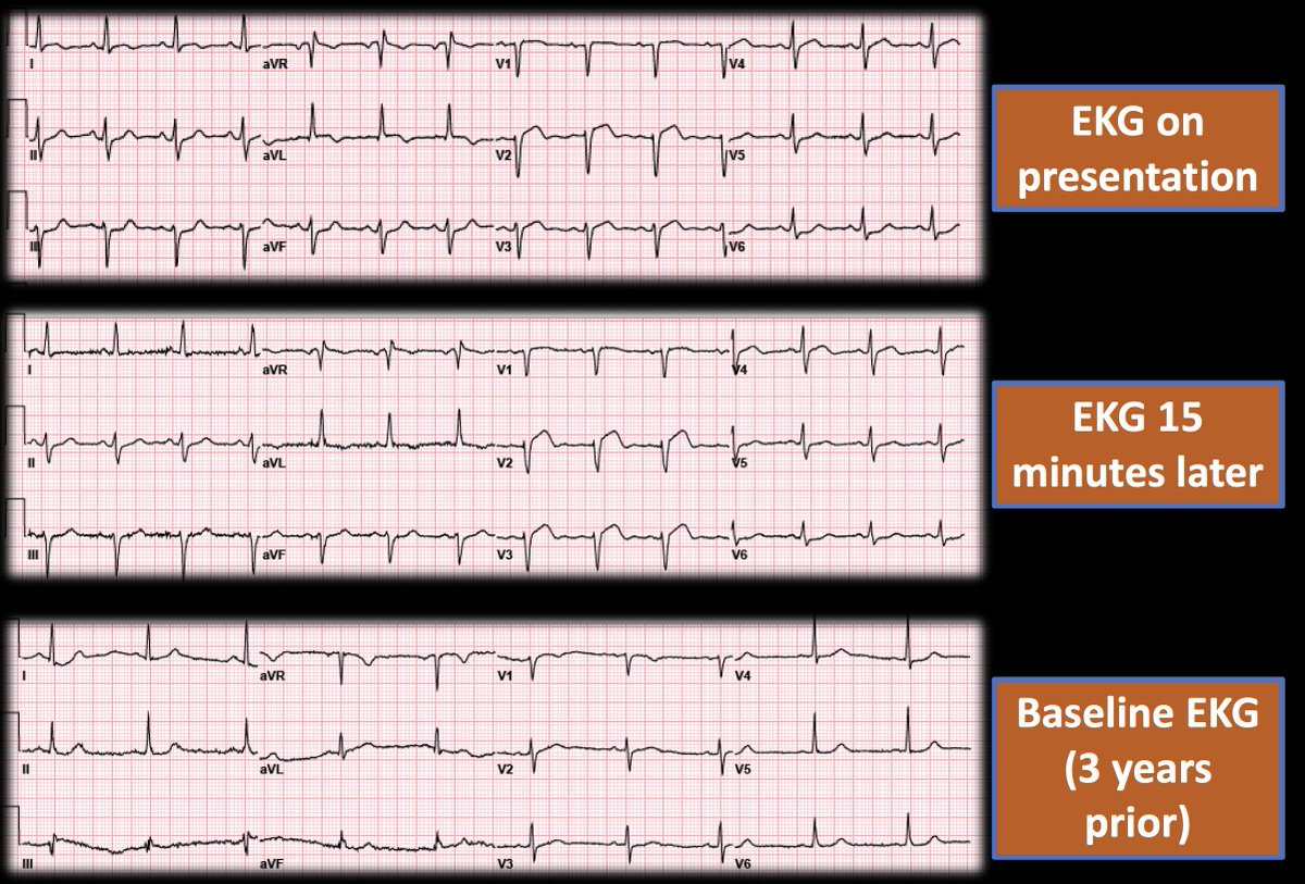
✨Interesting #EKG Case: Patient presented to ER with chest pain. Cath lab was activated after #ECG on presentation, but somehow got another EKG 15 min later 👇
❓What did the coronary angiogram show?🪡
#CardioTwitter #ACCFIT #Cardiology #MedTwitter BCM Cardiology Stephen W. Smith


Mechanism of Aortic Insufficiency? Is there a tear in the leaflet or dropout? 🤔
Young patient. Dilated LV (LVEDD 6.6 cm, LVEDV 250 cc). Asymptomatic. No known history of endocarditis.
#CardioTwitter #ACCFIT #Cardiology #EchoFirst American Society of Echocardiography BCM Cardiology Aga Khan University

Late presentation after Anterior #STEMI 💔
s/p Primary #PCI of large wrap-around LAD
Did well initially but on day 3, got hypotensive, with a new small pericardial effusion on #EchoFirst
Fellows: What happened? #YesCCT below 👇
#CardioTwitter #ACCFIT #Cardiology #MedTwitter

Tesfaye A. Telila MD, FACC,FSCAI. Fascinating case Tesfaye A. Telila MD, FACC,FSCAI.. Thanks for sharing!
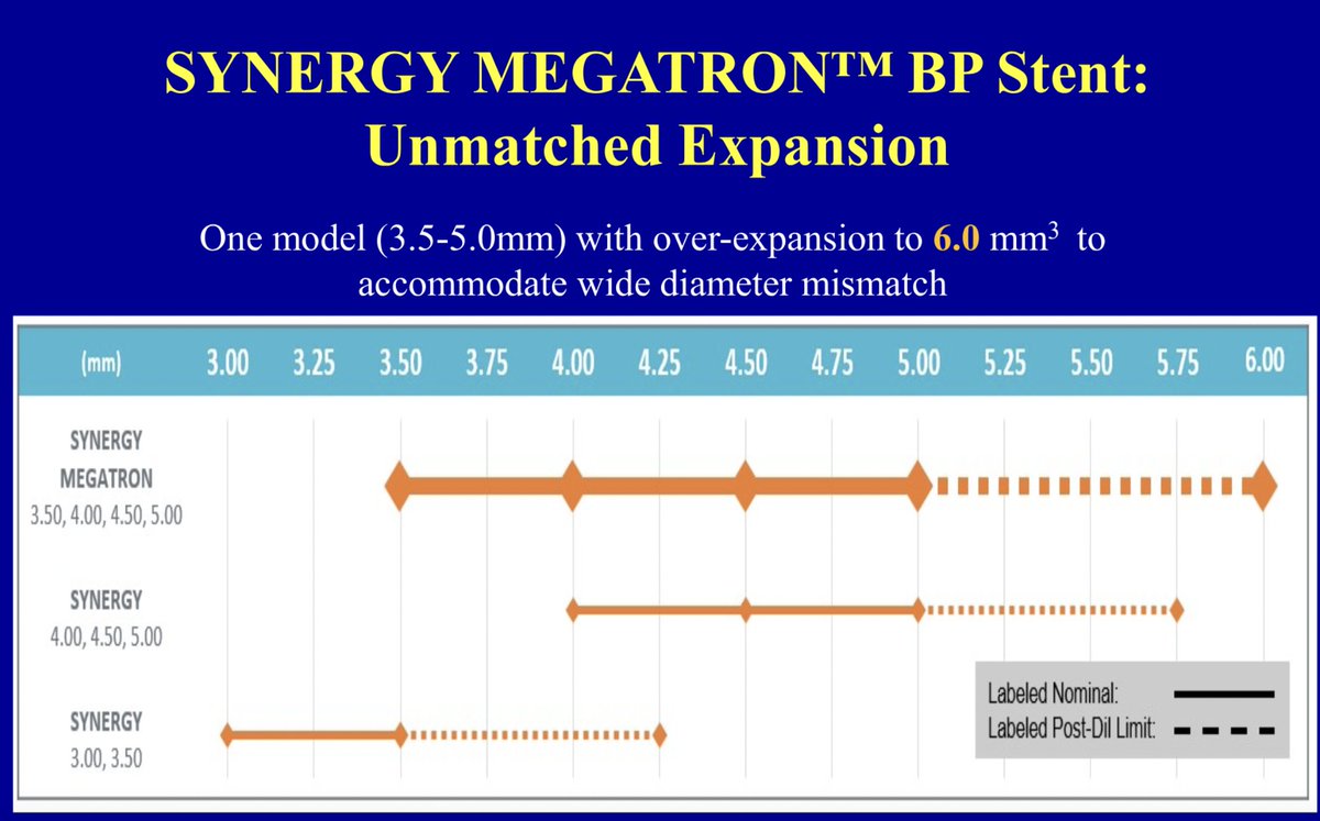
As others said, Megatron is the way to go here.
5.0 could be safely dilated to 6.0 without any disruption of polymeter/struts 👇

Dr Why jedicath աǟզǟʀ.ǟɦʍɛɖ Mamas A. Mamas Aditya Bharadwaj Poonam Velagapudi Jing Liu, MD Khalid Minhas, MD FACC Hooman Bakhshi Hady Lichaa, MD, FACC, FSCAI, FSVM, RPVI Antonious Attallah, MD, FACC, FSCAI Abdel Almanfi, MD, FACC, FSCAI Evandro Martins F. MD Here’s what we did:
1. RHC - confirmed his HF optimization, and guided in removal of #Impella @ the end.
2. Impella. Only spot was top of L femoral head. Loss of pulsatility on LM inflations.
3. Rota 1.5 LM-LAD. Shockwave LM-LAD.
4. IVUS-guided LM bifurcation stenting…

✨Ultra-Low Contrast PCI for bifurcation lesions: Case-based #Tweetorial 👇
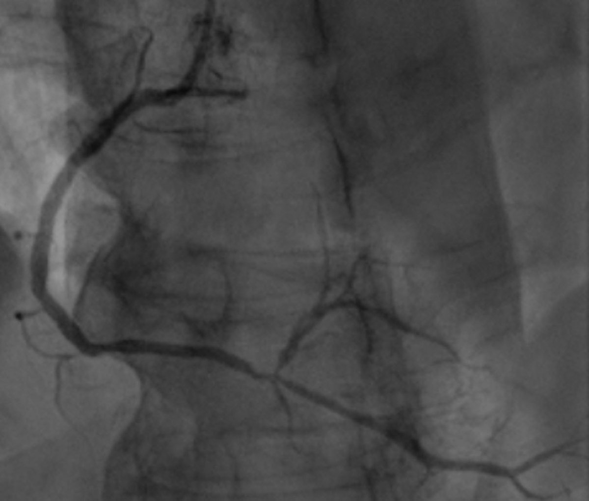
Diagnostic image below with lesion in RCA/rPDA/rPL bifurcation.
Creatinine = 3.1
Total contrast use = 3 cc (final cine)
1/10🧵
#CardioTwitter #ACCFIT #Cardiology #CardioEd

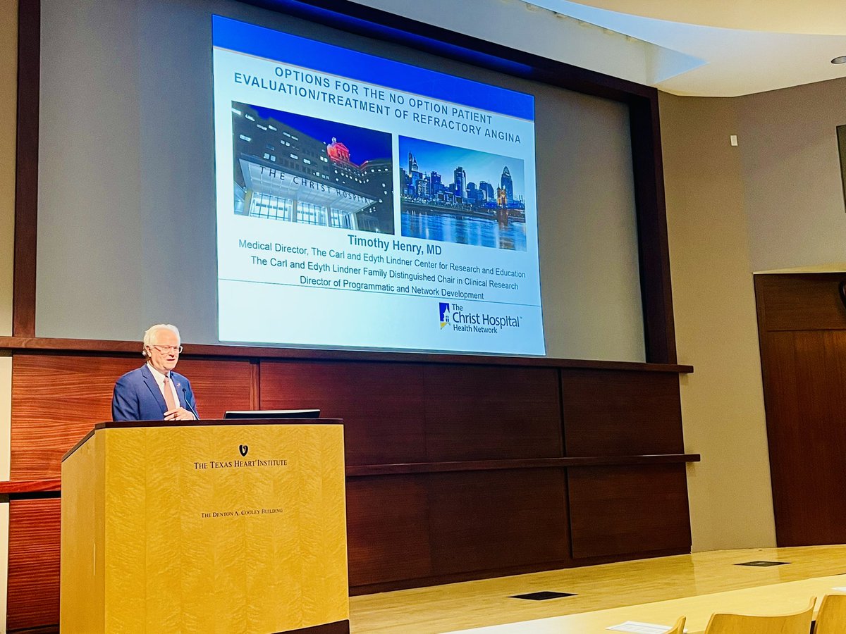
It was a pleasure to welcome Dr. Timothy Henry for 21st Annual John Lewis Interventional Lecture at Baylor College of Medicine / Texas Heart Institute. BCM Cardiology The Texas Heart Institute BCM Department of Medicine American Heart Texas Texas Chapter - ACC Texas Medical Center The Christ Hospital Health Network

🔹What will be ur approach for this #PCI ?
📌Transferred from OSH with this angio. Presented with HF now optimized.
📌82M, significant LMB stenosis, RCA #CTO , LVEF 10-15%, PAD. Turned down for #CABG
#CardioTwitter #Cardiology #ACCFIT European Bifurcation Club BCM Cardiology AKUCardiology

Qasim Raza, 𝘔𝘋 BCM Cardiology AKUCardiology American Society of Echocardiography American College of Cardiology Jing Liu, MD Benoy Shah MD Ozlem Bilen MD🫀FACC Anum Saeed Ritu Thamman MD Nicolas Merke Ambreen Mohamed, MD, FACC Jan Verwerft Hooman Bakhshi That's correct: Left bundle pacing wire. Appears to be more prominent that usual, but device interrogation was normal.
Sharing images below with location of HIS and Left Bundle pacing lead location on #EchoFirst (J Interv Card Electrophysiol (2022) 63:175–183) 👇 #EPeeps


What’s happening with the interventricular septum here 👀
History: Patient underwent uneventful BE-TAVR a day before. Doing well. No complaints. But then saw this on echo 😳
#CardioTwitter #Cardiology #ACCFIT #CardioEd #EchoFirst BCM Cardiology AKUCardiology American Society of Echocardiography American College of Cardiology


Waleed Kayani BCM Cardiology AKUCardiology Dr Shariq Shamim Mamas A. Mamas Hany Ragy Khalid Minhas, MD FACC Jing Liu, MD Aditya Bharadwaj Mahboob Alam Timir Paul, MD, PhD, FACC, FSCAI, FAHA Hady Lichaa, MD, FACC, FSCAI, FSVM, RPVI Nyal Borges G. Hanna J. Airton Arruda SREEVATSA NADIG Cardiologist DM FSCAI FESC Dr. Mukharjee Madivada Freddy_Dueñas MD Joy Sanyal,MD, DM, James Torey PA-C Swati Mukherjee Ahmad Alkonaiesy, MBBCh, M.Sc, MD, PhD 2/2 Here’s the angio. 3.5/38 stent, POT of LM w/ 4.5 NC as per OCT. Both diag & LCx were wired (provisional) and ok at the end.

Waleed Kayani BCM Cardiology AKUCardiology Dr Shariq Shamim Mamas A. Mamas Hany Ragy Khalid Minhas, MD FACC Jing Liu, MD Aditya Bharadwaj Mahboob Alam Timir Paul, MD, PhD, FACC, FSCAI, FAHA Hady Lichaa, MD, FACC, FSCAI, FSVM, RPVI 1/2 Here’s the OCT run from mid-LAD back into LM. pLAD had a fibrotic diffuse plaque extending back all the way to LAD ostium. This left us with 3 options:
i) nail the ostium (but not ideal coz diseased ostium ~ 2.5 mm within 4 mm LAD, undersized stent, risk of plaque shift)…

📌Two Qs regarding strategy for this #PCI ?
1. Ostial/proximal LAD appears to have a smaller lumen than mid LAD on angio. Where to land the stent proximally?
2. Diag - provisional?
#ACCFIT #CardioTwitter #Cardiology #CardioEd BCM Cardiology AKUCardiology

🚨 First reported case of #TAVR valvulitis possibly secondary to Pembrolizomab! Another critical consideration for ICIs? 🤔
Our superstar #ACCFIT BCM Cardiology, Azka Latif, presented this case at #ACC24 ! 🌟
#CardioTwitter #Cardiology #CardioEd #MedTwitter #cardioOncology 🫀


